Mucinous cystadenocarcinoma (↑ risk of perforation)
• Pseudomyxoma peritonei
Due to rupture of mucocele, usually malignant rather than benign mucocele
Peritoneal cavity filled with mucus
Loculated ascites; scalloped surface of liver and spleen
Envelopes and obstructs bowel
• Myxoglobulosis
Rare variant with multiple small globules ± calcifications
• Mucocele
Calcification (curvilinear) within wall or lumen
• Mucinous cystadenocarcinoma
Large irregular mass with thickened nodular wall
TOP DIFFERENTIAL DIAGNOSES
• Acute appendicitis (abscess)
• Appendiceal tumors
• Cecal carcinoma
• Ovarian cystic mass
• Cystic fibrosis
CLINICAL ISSUES
• Rare
• Complication: Rupture, torsion, intussusception
• Treatment
Surgical resection (usually right hemicolectomy)
• Prognosis
Simple mucocele and cystadenoma (good), carcinoma (poor)
– Pseudomyxoma: Poor prognosis
(Left) Axial CECT in a 30-year-old woman with chronic and acute RLQ pain shows an ileocolic intussusception , with a cystic-appearing lead mass that has calcification within its wall.
(Right) Another CT section in the same patient shows the intussusception and the mucocele that was the lead mass.
(Left) Gross pathology of the resected specimen in this case shows the intussusception and the mucocele .
(Right) Gross pathology in the same patient shows the resected specimen opened to illustrate the characteristic mucinous content of the mucocele.
TERMINOLOGY
Definitions
• Chronic cystic dilatation of appendiceal lumen by mucin accumulation
IMAGING
General Features
• Best diagnostic clue
Round or oval, thin-walled, cystic mass near tip of cecum
• Size
Usually 3-6 cm in diameter
• Other general features
Classified into 3 types based on histology
– Focal or diffuse mucosal hyperplasia
– Mucinous cystadenoma
– Mucinous cystadenocarcinoma
Focal or diffuse mucosal hyperplasia (simple mucocele)
– Resembles hyperplastic polyp of colon
– Does not perforate
Mucinous cystadenoma
– Benign neoplasm; most common type of mucocele
– < 20% of cases perforate with mucus seeding
Mucinous cystadenocarcinoma
– Less common than benign mucocele, but more likely to perforate and cause pseudomyxoma peritonei
Pseudomyxoma peritonei
– Due to rupture of mucocele, usually malignant
– Peritoneal cavity fills with mucus
Myxoglobulosis
– Rare variant with multiple small globules
– Calcify and produce 1-10 mm mobile calcifications
Only gold members can continue reading. Log In or Register to continue


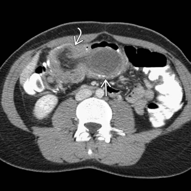
 , with a cystic-appearing lead mass
  that has calcification within its wall.
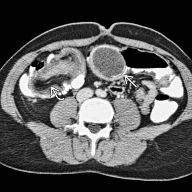
 and the mucocele
  that was the lead mass.
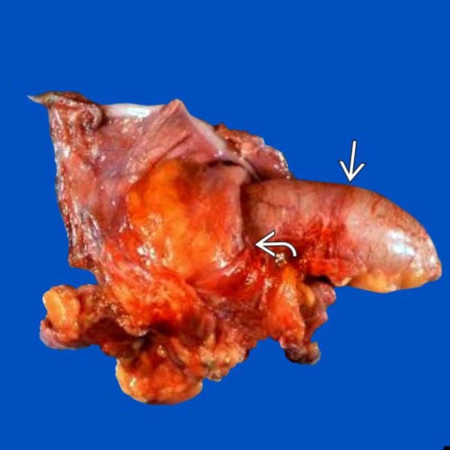
 and the mucocele
  .
.


 Myxoglobulosis


