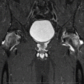
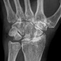
Risk factors for multifocal osteonecrosis
1. Corticosteroid use
2. SLE
3. Sickle-cell disease
4. HIV infection
5. Alcohol abuse
6. Multiple sclerosis
7. Coagulation disorders
8. Inflammatory bowel disease
9. Organ transplant recipients, e.g., renal, liver
10. Malignancy/chemotherapy
11. Sjogren’s syndrome/inflammatory arthritis
Although corticosteroid use is often reported as the most common risk factor in 90 % of patients with multifocal osteonecrosis, other associated conditions include renal failure, alcohol abuse, inflammatory bowel disease, systemic lupus erythematosus, human immunodeficiency virus infection, coagulation abnormalities (e.g., antithrombin III deficiency, protein S deficiency, factor V Leiden mutation, or increased activity of plasminogen activator), multiple sclerosis, Sjogren’s syndrome, sickle-cell disease, leukemia, and lymphoma. However, due to paucity of reports and a high incidence of asymptomatic lesions, our understanding of the true incidence of multifocal osteonecrosis in various individual disease conditions is limited. Nevertheless, one study on 200 patients with sickle-cell disease reported that multifocal involvement occurred in 44 % of patients (87 out 200 patients) [4]. While the underlying pathoetiology in the development appears to be multifactorial (e.g., venous hypertension, altered fat metabolism, mechanical stress, and primary cell death), recent reports suggest that familial thrombophilia, hypofibrinolysis, and genetic polymorphisms of the endothelial nitric oxide synthetase (e.g., 4a and T-786C) causing reduction in nitric oxide production may play a role [3].
Osteonecrosis is considered a well-recognized complication during the maintenance phase following treatment of acute lymphoblastic leukemia (ALL) and non-Hodgkin’s lymphoma. A higher incidence of lesions in the lower extremities compared to upper extremities with a predilection towards bilateral involvement. Multifocal involvement is also found to be more common in this population than previously reported, with 82 % of patients affected in one series who had undergone whole-body screening MRI following chemotherapy for acute lymphoblastic leukemia [5, 6]. This increase in incidence is potentially related to prolonged use of high-dose dexamethasone in the recent chemotherapeutic regimens and low threshold for ordering MRI scans in chemotherapy patients complaining of musculoskeletal symptoms. A complex interplay of pathogenetic factors such as suppression of bone formation, expansion of intramedullary adipocytes causing elevated intraosseous pressure, and secondary marrow ischemia has been postulated to predispose these patients to develop osteonecrosis. In a large retrospective study on 111 patients, Mattano et al. reported that a higher incidence of osteonecrosis was found in the age group between 10 and 20 years compared to younger children who were below 10 years of age [5]. This was potentially related to the improved buffering of the intraosseous pressure by the immature bone prior to the epiphyseal closure. In contrast to more recent reports, they found a 6 % incidence of multifocal osteonecrosis in their patients.
53.3 Location of Involvement and Clinical Features
Typically, when long bones are involved, it affects the epiphyseal region, although less commonly it can involve the metaphysis and the diaphysis [7]. In multifocal osteonecrosis, femoral head involvement is found to occur most commonly, followed by the knee, shoulder, and talus. Less common sites of involvement include the elbow (the trochlea and the capitellum), wrist (distal radius and other carpal metacarpal heads), and tarsal bones such as the cuboid, cuneiform, and the navicular bone. In two large reports on multifocal osteonecrosis, it was found that the mean number of skeletal sites involved was found to vary between 5.2 and 6.3 [2, 4]. Majority of the patients with multifocal disease present in the early pre-collapse stage of the disease (Ficat-Arlet stages 1 and 2), with either hip symptoms or multiple joint pain (Table 53.2).
Table 53.2
Patient demographics reported in studies on multifocal osteonecrosis
Author/study | No. of patients | Age/gender | Follow-up | Location in long bones | Diagnosis | Bones involved | Risk factors | Treatment |
|---|---|---|---|---|---|---|---|---|
Fajardo-Hermosillo et al. [8] | 1 | 24/W | 3.3 | Meta-diaphyseal | Multifocal ON | Tibia, fibula, talus | SLE associated with corticosteroids | NS |
Gonzalez-Garcia et al. [9] | 1 | 49/M | NS | Epiphyseal, meta-diaphyseal | Multifocal ON | Hip, knee, tibia, calcaneus, navicular, cuboid, metatarsals | HIV | NS |
Miettunen et al. [6] | 11 | 5.4 | NS | Epiphyseal, meta-diaphyseal | Multifocal ON | Distal femur, proximal tibia, ankle, proximal femur | ALL; post-chemotherapy | |
Sinclair et al. [7] | 1 | 42/F | NS | Meta-diaphyseal | Multifocal ON | Femur, tibia, talus | MS associated with corticosteroids | IM reaming |
Flouzat-Lachaniete et al. [4] | 49 | 46 | 15 | Epiphyseal, meta-diaphyseal | Multifocal ON | Hip, knee shoulder, and ankle | Sickle-cell disease | NS |
Solarino et al. [10] | 1 | 14 | 5.3 | Epiphyseal | Multifocal ON | Hip, knee, and shoulders | ALL, post-chemotherapy | Bilateral THA |
Gutierrez et al. [11] | 3 | NS | NS | Epiphyseal, meta-diaphyseal | Multifocal ON | Shoulder, hip, and knee | HIV | NS |
Mullan et al. [12] | 1 | 42/M | NS | Epiphyseal, meta-diaphyseal | Multifocal ON | Hip, talus, knee, shoulder | HIV | NS |
Collaborative study group [1] | 101 | 36 | NS | Epiphyseal, meta-diaphyseal | Multifocal ON | 6.2 lesions/patient hip, knee, shoulder, ankles, foot | Multiple factors | NS |
LaPorte et al. [2] | 32 | 34(24 women/8 men) | NS | Epiphyseal, meta-diaphyseal | Multifocal ON | Hip, knee, ankle, shoulder | Multiple factors | NS |

