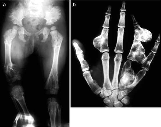
In Maffucci’s syndrome (very rare), multiple, diffuse chondromas are associated with multiple hemangiomas, cutaneous, subcutaneous, or in the deep soft tissues (not in the bone). Basic imaging is the same as described for solitary chondroma. In the metaphyses, longitudinal columns of radiolucency extend toward the diaphysis. They are divided by longitudinal bony septae, giving a characteristic W-like pattern. Chondromas can be very extensive, sometimes bubbly or trabeculated, with expansion of the bone, very thin cortex, or lack of any cortex. In Maffucci’s syndrome, phleboliths reveal on X-rays the angiomas. Histologically, compared to solitary enchondroma, lesions show features of more pronounced and persistent proliferative potential. Cartilage is more cellular and nuclei sometimes hyperchromatic, and the histology overlaps with low-grade chondrosarcoma by cytology alone. The stage is 2 in children, more frequently 1 in adults. Transformation to secondary sarcoma, most commonly chondrosarcoma, is frequent and probably ranges 20–30 % in Ollier and certainly higher in Maffucci (likely >50 %). Malignant transformation to sarcoma is usually seen in adults but may occur even before age 20, especially in Maffucci.
Both Ollier and Maffucci are conditions at increased risk to develop extra-skeletal malignancies, such as breast, liver, ovarian cancer and CNS tumors, suggesting an underlying genetic disorder predisposing to cancer in general. Surgical treatment is aimed to relieve symptoms rather than excise chondromas. Skeletal deformities and limb length discrepancy are addressed by osteotomies and/or lengthening procedures. Prognosis is burdened by the incidence of malignant change.
14.1 Concept of “Active” Chondromas and Differential Diagnosis with Chondrosarcoma
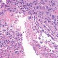
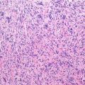
It is important to keep in mind that certain chondromas may show a histological pattern essentially similar to chondrosarcoma. In fact, multiple chondromas in Ollier and Maffucci, periosteal chondroma, enchondromas of the hands and feet, synovial chondromatosis, and soft tissue chondromas show histologic features consistent with grade 1 chondrosarcoma of the bone. In other words a grade 1 chondrosarcoma is cytologically indistinguishable from a benign lesion encountered in the abovementioned clinical settings. Therefore, diagnosis of malignant change is based on clinico-radiographic features and on the permeative growth pattern of tissue toward bone trabeculae. Secondarily, it is of paramount importance that the pathologist reviewing the slides has adequate clinical information, including site of biopsy and X-rays, and discusses the case with the orthopedic surgeon before making the diagnosis of chondrosarcoma. Quite important to the recognition of malignant change is to have a radiologic baseline taken in early adulthood (around age 20). Malignancy is characterized by a focal change in a bone of the baseline pattern often demonstrating a more “windblown” pattern often with a new soft tissue mass best seen on an MRI film. The patient also experiences chronic pain and may feel a new mass.