Case presentation
A 14-year-old male is brought to the Emergency Department after being involved in a motor vehicle crash. The patient was a restrained front seat passenger in a vehicle that was struck by another car while stopped at a red light.
The patient did not lose consciousness and has no complaint of head pain, neck pain, back pain, or abdominal pain. He states he feels that his right foot “was twisted” and “slammed into the floorboard” of the car. He complains of pain to the dorsal aspect of the foot. He denies numbness.
His physical examination reveals normal vital signs and is generally unremarkable. He has swelling and pain on palpation of the dorsal-lateral aspect of his right foot. There is no obvious deformity. The joint appears to be stable. He has brisk capillary refill and both the dorsalis pedis and posterior tibial pulses are equal and strong.
Imaging considerations
Plain radiography
This is the first-line imaging modality in patients with suspected bone injury. Patients with injuries that are suspected to involve the talus should have anterior-posterior, lateral, and oblique views of the ankle (an ankle series); if the talus head is not fully visualized, consideration should be given to obtaining an anterior-posterior view of the foot. Radiography has been shown to have difficulty in detecting talar dome fractures and lateral process fractures. ,
Computed tomography (CT)
This imaging modality is often used as a follow-up to plain radiography. CT can provide excellent bony detail, and if there is a complex fracture, the diagnosis of a talus fracture is in question, or there is strong clinical suspicion of a talus fracture with negative plain radiography, utilization of CT is appropriate. This is especially true as some talus fractures may be occult or not well depicted on plain radiographs, making CT useful in these cases. CT has been found to improve detection of talar dome fractures, improve talus fracture classification, and detect fractures of the lateral process of the talus, which can be difficult to visualize with plain radiography. ,
Magnetic resonance imaging (MRI)
This is not a first-line imaging modality for the evaluation of fractures but can be useful when there are significant symptoms and a strong clinical suspicion of injury with negative plain radiography or equivocal CT. MRI is especially useful for the evaluation for osteochondral injury (e.g., talar dome osteochondral injury), ligamentous injury, acute or chronic physeal injury, and stress fracture.
Imaging findings
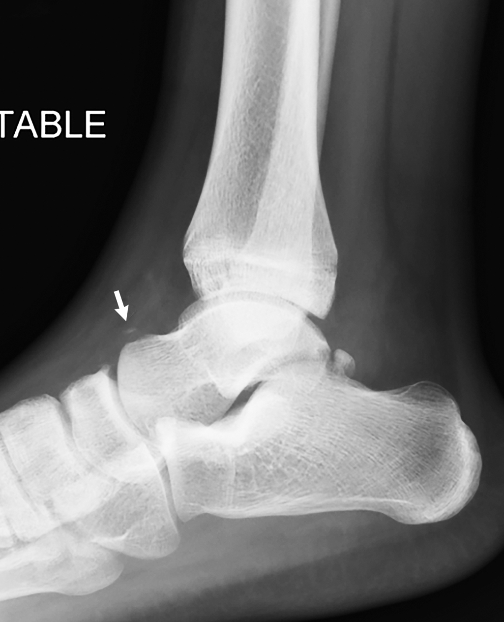
Three-view plain radiography of the right ankle (anterior-posterior, lateral, and mortise views) was obtained. These images demonstrate a thin, curvilinear ossification dorsolateral to the distal talar neck consistent with an acute avulsion chip fracture of the talus neck. The ankle mortise is normal in appearance ( Figs. 69.1–69.3 ).


Case conclusion
Pediatric Orthopedics was consulted. The patient was placed in an ankle boot, given crutches, and advised to be non–weight bearing until follow-up with Pediatric Orthopedics within 1 week of discharge from the Emergency Department. The injury healed well and the patient did not develop complications from his injury.
Fractures of the talus are not common and are the result of an axial loading injury to the anterior portion of the tibia with the foot in dorsiflexion. The reported incidence of talus fracture in the pediatric population is 0.1%–0.8%. , Previously reported etiologies causing talus fractures include motor vehicle accidents and falls from significant heights. , The neck of the talus is the most common fracture site, followed by the body.
Talar neck fractures represent 50% of all talus fractures and are well described in the litertature. Talar body fractures are the second most common fracture of the talus and are seen in high-energy mechanism injuries and activities that involve landing on one’s feet (e.g., cheerleading and gymnastics injuries) resulting in shear fractures of the talar body. Fractures of the talar head are uncommon and there is sparse literature on this injury. Less than 10% of talus fractures involve the head, and outcomes are not well documented; often, this fracture is associated with other fractures, typically from high-energy injuries. A unique fracture is a lateral process fracture, also known as “snowboarder’s fracture” since this fracture is more common in participants of this sport; the injury occurs when the foot is in a dorsiflexed and inverted position. These fractures are frequently missed. ,
Talus fractures have associated complications, including avascular necrosis (AVN) (7%), arthrosis (17%), delayed union (3%), neurapraxia (7%), and the need for further surgery (10%). These complications have been found to be more common in adolescent boys. , , AVN is more commonly associated with fracture-dislocations, and while the exact mechanism that leads to the development of AVN is not well understood, dislocation is a risk factor. , One study found that older patients (above the age of 12 years) had a higher incidence of fracture-dislocations, which were more likely to be associated with vascular injury. Another study found low rates of AVN, but like previously reported series AVN was seen in patients with displaced fractures. This older group also was more likely to require operative intervention. AVN has been reported in patients with minimally displaced fractures, so prompt follow-up and monitoring are important for patients with fracture-dislocation injuries. , A review of reported talus fracture series since 1985 in pediatric patients found an incidence of AVN of 16% in 83 cases of nondisplaced talus fractures and 51% in 49 cases of displaced and comminuted talus fractures.
The Hawkins Classification System has been used to describe talus fractures :
| TYPE | DESCRIPTION |
| I | Talar neck fracture with no to minimal displacement |
| II | Talar neck fracture with subtalar dislocation |
| III | Talar neck fracture with subtalar and tibiotalar dislocations |
| IV | Talar neck fracture with subtalar, tibiotalar, and talonavicular dislocations. |
Related posts:


Stay updated, free articles. Join our Telegram channel

Full access? Get Clinical Tree


