Introduction
The concept of injecting a tracer into the blood stream and detecting its transit and distribution in the heart muscle for the assessment of myocardial perfusion is well established in nuclear cardiology and cardiovascular magnetic resonance (CMR). Both exogenous, injected contrast agents and endogenous contrast mechanisms have been used to assess myocardial perfusion with CMR. The use of a gadolinium (Gd)-based contrast agents for the assessment of myocardial perfusion with CMR has been extensively validated and successfully applied in patient studies (see Chapter 18 ). Recent developments, in particular the introduction of parallel imaging, and sparse sampling methods (see Chapter 5 ), have made it possible to combine the requirements for spatial and temporal resolution for myocardial perfusion imaging during the first pass, with multi-slice coverage of the heart. The need for a quantitative, largely observer-independent analysis of perfusion studies has also received increasing support. Several approaches have been developed that allow quantification of myocardial blood flow (MBF) with CMR. Advances in rapid imaging techniques for CMR perfusion imaging (covered in detail in Chapter 5 ) and the development of methods to obtain reproducible, quantitative measures of myocardial perfusion have established CMR perfusion imaging as an attractive alternative to nuclear perfusion imaging.
The Physiologic Basis for Measuring Myocardial Perfusion
Coronary circulation blood flow resistance is under normal conditions determined primarily by the myocardial microcirculation, vessels with a diameter less than 300 µm. The adequate supply of oxygen and metabolites to the myocytes is tightly coupled to MBF. The assessment of microcirculatory function has therefore been used as a surrogate marker for the detection of myocardial ischemia, although ischemic conditions are also critically dependent on the workload and metabolic function. For example, under conditions of myocardial hibernation, the degree of ischemia is reduced by a corresponding reduction of the cardiac workload. Adequate and near-constant blood flow is maintained through autoregulation of coronary tone. Compensatory vasodilation can maintain adequate myocardial blood under resting conditions for up to 80% diameter epicardial coronary artery narrowing. With more severe narrowing in an epicardial vessel, and in the absence of significant collateral flow, the distal perfusion bed is fully vasodilated even under resting conditions, and no augmentation of blood flow is feasible during periods of increased demand (e.g., if the patient exercises or undergoes pharmacologically induced stress). In healthy subjects, MBF can increase 3- to 4-fold with maximal vasodilation. This means that differences in MBF between a region subtended by a stenosed coronary artery and the territory of a normal coronary artery are significantly amplified with maximal vasodilation.
Myocardial perfusion imaging during pharmacologic vasodilation (e.g., with adenosine, dipyridamole, or regadenoson) rests on the physiologic observation that the hemodynamic significance of an epicardial lesion is most apparent during maximal vasodilation. A related measure can be obtained in the catheterization laboratory with an intravascular Doppler flow probe by measuring the coronary flow reserve, to assess lesion severity, or by measuring the fractional flow reserve, defined as mean distal coronary artery pressure divided by the aortic pressure during maximal vasodilatation. These functional tests of coronary resistance and MBF overcome the known limitations of vessel lumen diameter measurements by x-ray angiography for determining the hemodynamic significance of epicardial lesions.
Myocardial perfusion imaging can also be used to assess functional impairments in the coronary microcirculation, when there are no epicardial lesions. Both myocardial perfusion reserve, and coronary flow reserve were found to be abnormally low in women with microvascular dysfunction, and without significant epicardial stenoses. There is also evidence from epidemiologic studies that it is feasible with CMR perfusion imaging to detect subclinical disease and silent ischemia in persons without a history or symptoms of atherosclerotic disease. The myocardial perfusion reserve in response to adenosine was found to be associated with coronary artery disease risk factors in patients with a low likelihood of significant epicardial disease. In asymptomatic patients a lower myocardial perfusion reserve is associated with decreased regional left ventricular (LV) function and also with lower regional myocardial strain.
With the development of CMR perfusion imaging, it has become possible to probe with sufficient spatial resolution for more subtle indicators of myocardial ischemia. Blood flow across the myocardial wall is not uniform but instead favors the subendocardium to accommodate the higher workload and higher rate of oxygen consumption in the subendocardial layer. Under normal resting conditions the ratio of endocardial to epicardial blood flow is on the order of 1.15 : 1. With a coronary artery stenosis, blood flow is first diverted away from the subendocardial layer and the endocardial-to-epicardial blood flow ratio is often <1 : 1, in particular when under stress. With myocardial ischemia, the subendocardial layer is accordingly most susceptible to necrosis. The transmural gradient of MBF can only be sufficiently appreciated if the imaging modality provides spatial resolution on the order of ≤2 mm. With CMR it has become feasible to detect flow impairments limited or most accentuated in the subendocardial layer. The spatial resolution of conventional imaging modalities such as positron emission tomography (PET) and single photon emission computed tomography (SPECT) was insufficient to detect MBF deficits limited to the subendocardial layer. More specifically, the sensitivity of a myocardial perfusion imaging technique is directly related to its spatial resolution, and the ability to discern transmural variations of flow.
CMR offers the unique possibility of quantitatively assessing both perfusion, viability, and function with high accuracy, to distinguish, stunned, hibernating, and infarcted myocardium. Bolli et al. showed that even small differences in blood flow during ischemia result in large differences in postischemic function, suggesting that the ability to quantify flow in the low-flow range is of importance to predict the probability of postischemic recovery. The extent and incidence of microvascular obstruction observed with CMR were associated with the duration of ischemia before coronary intervention.
First-Pass Imaging With Exogenous Tracers
The success of applying CMR to detect the first pass of an injected contrast agent and detect perfusion abnormalities starts with an understanding of the contrast mechanisms that allow contrast agent detection. The first-pass transit of a contrast agent through the vasculature and tissue leads to changes in the T1 and T2* relaxation time constants for the detected 1 H signal: the contrast reagent is not detected directly, but rather through its effects on the signal from 1 H nuclei in tissue. The chelates of paramagnetic Gd ions used as contrast reagents have a relatively high magnetic susceptibility, which causes on a microscopic scale magnetic field inhomogeneities and shortens the transverse (T2*) relaxation rate of water (see Chapter 3 ). The contrast reagent also shortens the longitudinal (T1) magnetization recovery after a radiofrequency pulse has disturbed the equilibrium state.
CMR methods for perfusion imaging can be subdivided into T1- and T2*-weighted techniques: T1-weighted techniques produce signal enhancement during the transit of the contrast agent, whereas T2* techniques cause signal loss. For contrast agents confined to the vascular bed, and in the absence of significant organ motion, T2*-weighted perfusion imaging gives rise to relatively larger signal changes than T1-weighted techniques, because the magnetic susceptibility effects of the contrast agents extend beyond the capillaries. But T2*-weighted imaging techniques have an inherent sensitivity to motion, leading to signal loss in the presence of motion. For myocardial perfusion imaging, T1-weighted techniques such as gradient echo imaging with short echo times, and a magnetization preparation for optimal T1 weighting have therefore steadily gained preference in CMR. We provide here a brief overview of three major sequence techniques for T1-weighted perfusion imaging of the heart. In historical order, they are spoiled gradient echo imaging (e.g., “TurboFLASH” or “turbo field echo” [TFE]); gradient echo imaging with balanced steady-state free precession (bSSFP); and multi-shot T1-weighted echo planar imaging.
Single-shot gradient echo imaging with a magnetization preparation (e.g., TurboFLASH or TFE) is well suited for T1-weighted, quantitative myocardial perfusion imaging. A magnetization preparation for T1 weighting can take the form of an inversion pulse or a saturation pulse; either of them is generally applied in a non-slice-selective mode so that blood flowing into the image plane during the subsequent image acquisition has been subjected to the same T1 preparation as myocardium. In a sequential multi-slice imaging sequence, a saturation preparation can be repeated for each slice, and this will result in the same degree of T1 weighting for each slice. After the magnetization preparation, images for one or more slices are rapidly read out within approximately <200 ms per image, or even less depending on the imaging acceleration technique that is being used. Typical sequence parameters for such rapid readouts are a repetition time per phase-encoding step (TR) of ≤2.5 ms, an echo time (TE) of ≤1 ms, a receiver bandwidth on the order of 400 to 700 Hz/pixel, and an in-plane spatial resolution of 2 to 3 mm. Because of the short TE, the signal is relatively insensitive to flow and magnetic susceptibility variations (e.g., between tissue and blood pool during the first pass). With a linear ordering for the phase-encoding steps, the image acquisition only needs to be delayed by 10 to 20 ms after a saturation recovery preparation. Depending on the effective time after saturation (TS) or time after inversion (TI, if an inversion pulse is used), the signal increases approximately linearly with contrast agent concentration, or as a function of the relaxation rate constant. (Note that the relaxivity of the contrast reagent is not appreciably different between blood and tissue.) Eventually the signal ceases to increase because of the opposite effect of T2* on the signal at higher contrast concentrations, and even in the case of negligible TEs (e.g., with image readouts with ultra-short TE) the signal intensity from a proton density–weighted image represents an effective upper limit for the signal intensity dynamic range. Fig. 4.1 shows an example of a CMR perfusion study with a spoiled gradient echo technique.

The signal-to-noise (SNR) and the contrast-to-noise ratio (CNR) with gradient echo perfusion imaging are relatively low, when short TRs, and wide receiver bandwidths are used. The maximum signal intensity is reached with relatively low flip angles. (The flip angle corresponding to maximum signal intensity is referred to as “Ernst angle.”) To overcome this limitation, one can use a bSSFP image readout, which maximizes the preservation of phase coherence of the transverse magnetization between repetition periods (TRs) in the pulse sequence, and efficiently converts magnetization between transverse and longitudinal orientations. With bSSFP, the Ernst angle can approach 90 degrees—that is, one can reach the maximum theoretical signal amplitude after each radiofrequency (RF) excitation, while still repeating the excitations with very short TRs. In fact, the TR should be as short as feasible, because any off-resonance phase shifts increase signal loss, and phase shifts increase with TR, and also with flow. Off-resonance frequency shifts result in dark bands at locations where the frequency shift (in radians/s) times TR (in seconds) equals an odd multiple of π/2.
The passage of a contrast bolus in the LV cavity can produce a frequency shift as large as approximately 100 Hz at 1.5 T and larger at higher field strengths. As the off-resonance shift varies with changing contrast loading of the blood pool during the first pass, it can induce signal variations over the myocardium, which can mimic perfusion defects (not to be mistaken with dark-rim artifacts, which are discussed later and which have a different appearance). At field strengths of ≥3 T, bSSFP image readouts during first-pass imaging can give rise to quite severe image artifacts, with dark bands appearing over the myocardium. The use of bSSFP readouts has therefore been mostly limited for cardiac perfusion imaging to field strengths of ≤1.5 T. Although the bSSFP readouts can produce appealing image quality, its use has to be approached with caution as the artifacts on bSSFP images can be deceptive.
Echo planer imaging (EPI) eliminates the need for RF excitation before each phase-encoding step, and, as a result, it is one of the fastest imaging methods for freezing heart motion (50–100 ms for single-shot acquisitions). In an EPI pulse sequence, an initial RF excitation creates a coherent precession of transverse magnetization, and a train of gradient echoes is then generated by applying a rapidly oscillating magnetic field gradient along the readout direction. Each of these gradient echoes is preceded by a short, blipped gradient pulse, applied along a direction perpendicular to the oscillating magnetic field gradient. The blipped phase-encoding gradients between echoes advance the trajectory in the frequency acquisition space ( k -space) perpendicular to the readout direction. Instead of just a single line, one acquires after each RF excitation a set of parallel lines in frequency space (“ k -space”). The T2*-related decay of the gradient echo amplitudes in the echo train results in an increasing sensitivity of later echoes to T2* and motion and causes blurring. The effective TE can be an order of magnitude longer for EPI sequences compared with fast gradient echo sequences (e.g., 30–40 ms vs. 1.2–2.0 ms for fast gradient echo imaging). Unfortunately, a longer effective TE gives rise to magnetic susceptibility and flow artifacts in the LV cavity. Furthermore, flow- and susceptibility-related artifacts in the LV make it difficult to measure the arterial transit of the contrast agent. To partially capture the speed advantage of EPI, and to minimize T2*- and flow-related artifacts, one reduces the length of the echo trains, and the images are read out with several RF excitations/echo trains. This results in a 30% to 40% speed advantage compared with conventional gradient echo sequences, and is particularly useful at field strengths of ≤1.5 T. (At ≥3 T, any increase of echo time beyond the minimum allowed by the gradient system nearly inevitably gives rise to susceptibility artifacts, in particular during passage of a contrast bolus.)
Ultra-fast T1 measurements ( Fig. 4.2 ) would provide the most accurate estimates of contrast agent concentration, and, with the advent of parallel imaging, this approach may become practical. Chen et al. developed such a T1 Fast Acquisition Relaxation Mapping (T1-FARM) method to obtain single-slice T1 maps of the heart with 1 s resolution. With a direct T1 measurement, the problems associated with the saturation of the signal with increasing contrast agent dosage can be avoided. This should allow for a more accurate quantification of blood flow using higher contrast agent dosages. A T1-based estimation of the contrast concentration is particularly advantageous for the blood pool, as saturation of the contrast enhancement is of foremost concern for the arterial input. If instead one measures T1 in the blood pool during the first pass of the contrast agent, the arterial input can be reconstructed from these T1 data rather than the signal intensity because the latter suffers from saturation effects. Chen et al. developed a slice-interleaved radial imaging technique, that runs continuously and can be used for reconstruction of cardiac phase-resolved images, and beat-by-beat estimation of T1 for arterial input sampling.

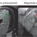
In its current state, all CMR techniques offer higher spatial resolution than is feasible with nuclear imaging techniques. An illustration of this is shown in Fig. 4.3 from studies in an experimental animal model.

Advanced Techniques for Perfusion Imaging Acceleration
Myocardial perfusion CMR has been at the forefront of developments in image acceleration techniques aimed at increasing coverage of the LV. Increased coverage can translate into a larger number of image slices, or, by switching to three-dimensional image acquisitions, increase spatial coverage of the myocardium. The increased LV coverage should not come at the cost of lower temporal resolution—for example, by increasing the repetition time for each slice location from one to two heart beats—because a lower temporal resolution will affect the accuracy for distinguishing gradual changes in MBF, in particular during stress conditions. Chapter 5 of this book is dedicated to advanced techniques for CMR perfusion imaging that provide increased coverage of the LV, possibly higher in-plane spatial resolution, and suffer from fewer image artifacts (e.g., “dark-rim” artifact). This chapter will instead focus on the underlying theory and principles of CMR myocardial perfusion imaging.
Endogenous Contrast for the Assessment of Myocardial Perfusion
Approaches have been developed for an assessment of myocardial perfusion that are not based on the use of an exogenous MR contrast agent but instead exploit endogenous contrast mechanisms related to blood flow and blood oxygenation. These approaches fall under the categories of spin labeling, blood-oxygen-level-dependent (BOLD) contrast, and magnetization transfer (MT) contrast. These techniques have to be considered technically more challenging than measurements with exogenous contrast agents, or offer only an indirect measure of blood flow.
With spin labeling, the spins are either inverted or saturated in a slab that is generally located upstream from the imaged slice. The flow-dependent change of signal intensity because of the inflow of saturated or inverted spins into the image slice provides a measure of tissue perfusion. The spin-labeling method generally relies on the assumption that the net arterial blood flow in the myocardium follows the direction from base to apex. Cardiac motion complicates the interpretation of signal changes in a spin-labeling experiment because the labeled spins can be transported into the imaged slice either through blood flow or by through-plane motion of the heart.
BOLD offers a measure of hemoglobin saturation reflecting regional oxygen supply and demand. Deoxyhemoglobin is paramagnetic, whereas oxyhemoglobin is only diamagnetic, which means that deoxyhemoglobin causes a considerably larger reduction of T2* than oxyhemoglobin and therefore a larger signal intensity attenuation in gradient echo images. Deoxyhemoglobin can be used as an endogenous intravascular tracer because of the tight coupling between oxygen demand and blood flow. BOLD contrast changes have been observed in the heart after administration of dipyridamole and dobutamine. The link between BOLD contrast and blood flow depends on the balance between blood flow and oxygen metabolism—that is, between oxygen supply and demand.
Spin labeling, BOLD, and MT contrast CMR have an advantage in that they can be carried out in the steady state, meaning there is no need to capture with ultra-fast imaging the transient signal changes observable in the myocardium after injection of an exogenous tracer. Ultimately these techniques may allow an assessment of myocardial perfusion with very high spatial resolution (~1 mm or less).
Quantitative Evaluation of Myocardial Perfusion
The images acquired in a “first-pass” study of myocardial perfusion—meaning rapid dynamic imaging during the first pass of an injected contrast bolus—can be qualitatively evaluated for differential signal enhancement in the myocardium. With a T1-weighted imaging technique, myocardial segments showing reduced signal intensity enhancement during the first pass, relative to other myocardial segments, are interpreted as hypoperfused. This type of qualitative judgment of signal enhancement differences can suffer from substantial observer bias and small reductions in perfusion may be missed. Although higher dosages of contrast agent would increase the dynamic contrast enhancement, and thereby render myocardial regions with reduced contrast enhancement more apparent, the images are also more likely to be contaminated by artifacts near the endocardial border. Furthermore, global reductions of blood flow because of diffuse microvascular ischemia will be missed by this approach. It then becomes advantageous to evaluate the contrast state during stress or vasodilation, relative to a baseline or resting state to uncover global impairments of perfusion.
With a fast T1-weighted gradient echo sequence and low contrast agent dosages (e.g., 0.03 mmol/kg of Gd-DTPA for an intravenous bolus injection into an antecubital vein) the myocardial contrast enhancement is approximately proportional to the local contrast agent concentration. Under these conditions the signal curves can be interpreted as, or transformed into, contrast agent residue curves. Absolute units do not matter here as much as a faithful reproduction of the relation in contrast concentrations between tissue and blood pool—enhancement in the tissue is interpreted relative to the enhancement in the blood pool. In fact, the tissue is often, for the purpose of quantifying perfusion, viewed as a linear, stationary system, where the enhancement in the tissue can be modeled as a linear response to arterial input of contrast.
Investigators using PET have over the years presented compelling arguments for a quantitative approach to myocardial perfusion imaging, including absolute quantification of MBF. With CMR, a quantitative analysis of contrast enhancement is based on signal intensity curves, which can be generated for user-defined regions of interest (ROI), or at the pixel level. CMR perfusion studies require a careful approach for image segmentation and registration. CMR perfusion studies are not motion-averaged, the endocardial borders are relatively well defined during transit of contrast through the ventricular cavity, and the large signal enhancement in the ventricular cavity requires accurate image segmentation to avoid spill-over effects. Fig. 4.4 shows an example of a CMR perfusion study in a patient for absolute quantification based on model-independent analysis of signal intensity curves.

In the vein of Zierler’s original work on the central volume principle, we consider here a tissue ROI with a single arterial input and a single venous output. The injection of a tracer is described by the variation of tracer concentration at the (arterial) input c in (t) . The amount of tracer that remains in the tissue region at any time t , q(t) , represents the difference between the amount supplied to the tissue region up to time t, and the total amount of tracer that has exited the region of interest up to the time t :
q(t)=F∫0t[cin(s)−cout(s)]⋅dt
Related posts:


Stay updated, free articles. Join our Telegram channel

Full access? Get Clinical Tree


