1. Collapse of the femoral head without joint-space narrowing or acetabular abnormality on x-rays (including crescent sign)
2. Demarcating sclerosis in the femoral head without joint-space narrowing or acetabular abnormality
3. “Cold in hot” on bone scans
4. Low-intensity band on T1-weighted images (band-like pattern)
5. Trabecular and marrow necrosis on histology
26.3 JIC Classification
Massive collapse of the femoral head is a crucial event that leads to the functional devastation of the hip joint with ONFH. To evaluate the susceptibility for collapse or to predict impending collapse for each case in the early stage of ONFH, the JIC proposed a classification in 1986 [1]. It consisted of three major types, based on the following specific radiographic findings (Fig. 26.1): type 1, demarcating sclerosis; type 2, flattening; and type 3, cystic radiolucency. Type 1 and type 3 are divided into subtypes 1A, 1B, 1C, 3A, and 3B, based on the location of the lesion in the weight-bearing area. A type 3A cystic lesion is located far from the weight-bearing surface. A lesion surrounded by demarcating sclerosis and located far from the weight-bearing surface is classified as type 1A. The prevalence of collapse was reported as 0 % in type 1A, 44 % in type 1B, 88 % in type 1C, 100 % in type 2, 0 % in type 3A, and 100 % in type 3B [4, 5]. Surgical treatment is seldom necessary in type 1A and type 3A. Varus femoral osteotomy or transtrochanteric rotational femoral osteotomy can be good indication for type 1B and 1C. The necrotic lesions were not able to be quantitatively evaluated on radiographs in type 2 and type 3 ONFH because they did not show demarcating sclerosis until the late stage. These types were often seen in patients with steroid-induced ONFH. Failure rates of osteotomies for type 2 and type 3B are high. The location of the lesion in these types was shown to occupy all weight-bearing area when MRI was used [2]. Therefore, a high prevalence of collapse in type 2 and 3B can be explained by the location of the lesion that occupied the most of the weight-bearing area of the femoral head.
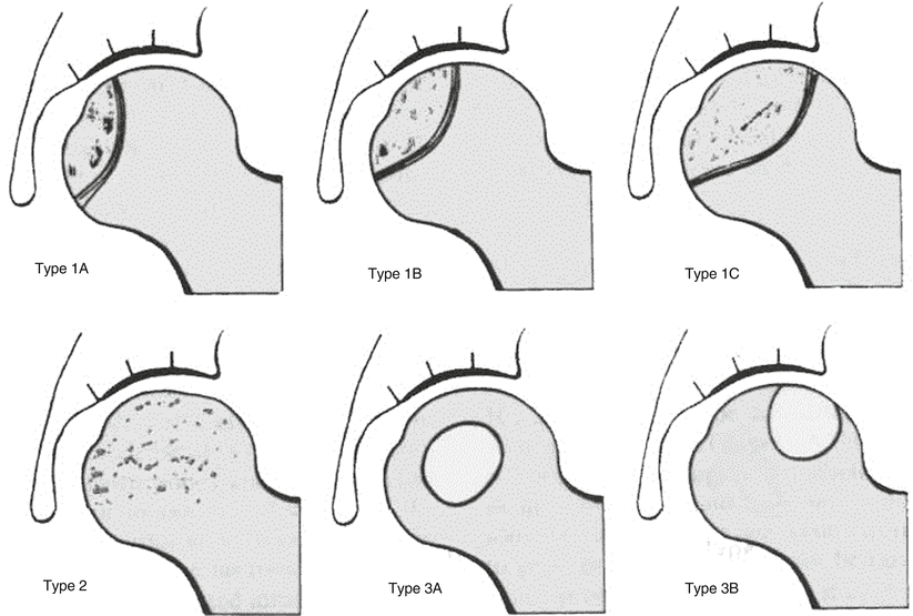
Fig. 26.1
The JIC 1986 radiographic classification of ONFH. The classification scheme consists of three types (1, 2, and 3). Type 1 is characterized by the presence of a demarcation line in the femoral head and is divided into three subtypes, 1-A, 1-B, and 1-C, according to its relationship to the weight-bearing surface. Type 2 shows early flattening of the weight-bearing surface but has no demarcation line around the necrotic area. Type 3 has cystic lesions and is divided into two subtypes according to their site in the femoral head
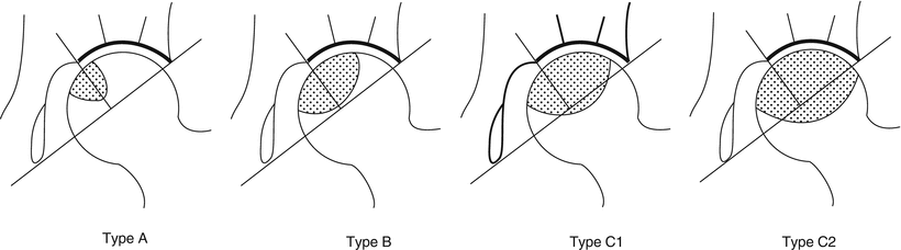
As the location of lesion is an important factor to predict impending collapse, a prospective MRI study in patients with SLE was conducted to detect ONFH in the pre-radiographic stage and to prognosticate ONFH [6]. A low-intensity band in the femoral head of normal fat intensity on T1-weighted images was a specific finding of ONFH. The lesions demarcated by a low-intensity band were classified into type A, B, and C like type 1A, 1B, and 1C of the radiographic classification on the coronal image through the femoral head center. As the location of the lesion extended more weight-bearing area, the prevalence of collapse increased. Although some early lesions detected less than a year after initial steroid treatment can show size reduction, the location of lesions does not change later in most of the cases [7, 8]. The MRI classification system based on the location of the lesion surrounded by a low-intensity band on T1-weighted images was found to have the same predictive power as the radiographic classification system. Furthermore, when type C was divided into subtypes C1 and C2, the incidence of progressive collapse of the femoral head varies significantly. A type C2 lesion extends laterally to the acetabular edge, whereas a type C1 lesion does not (Fig. 26.2). The prevalence of collapse was less than 10 % in type A, 40 % in type B, 80 % in type C1, and 90 % or more in type C2. Even after collapse occurs in type A and B, subsequent cessation of collapse can be expected and improvement of symptoms with no surgical intervention required [9]. This new classification was adopted in the 2001 JIC classification and it has not changed since then.
 Ischemia and Reperfusion Injury in Bone
 Glucocorticoids (as an Etiologic Factor)
 Alumina Ceramic-on-Highly Cross-Linked Polyethylene Bearing in Cementless Total Hip Arthroplasty in Young Patients with Osteonecrosis of Femoral Head
 Impaction Bone Grafting
 Causes of Pain in Early and Advanced Stage Osteonecrosis
 Kienböck’s Disease of the Lunate


Related posts:
Ischemia and Reperfusion Injury in Bone
Glucocorticoids (as an Etiologic Factor)
Alumina Ceramic-on-Highly Cross-Linked Polyethylene Bearing in Cementless Total Hip Arthroplasty in Young Patients with Osteonecrosis of Femoral Head
Impaction Bone Grafting
Causes of Pain in Early and Advanced Stage Osteonecrosis
Kienböck’s Disease of the Lunate
Stay updated, free articles. Join our Telegram channel
Full access? Get Clinical Tree
