Figure 7.1.1
Normal nuchal translucency. Sagittal image of the fetus in neutral position, magnified such that the fetus takes up more than half the image, shows a thin hypoechoic band (calipers) along the back of the fetal neck. The calipers have been placed to measure the thickness of the hypoechoic band, just inside the bordering tissues.
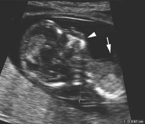
Figure 7.1.2
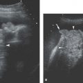
Abnormal nuchal translucency in hyperflexed fetus. Sagittal image of fetus with abnormally thickened nuchal translucency (calipers, 6.2 mm). The image is suboptimal for measuring the nuchal translucency, because the fetal neck is hyperextended, with a greater than 90-degree angle between the chin (arrowhead) and the chest (arrow).
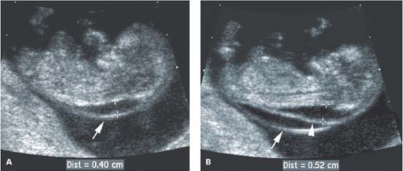
 Figure 7.1.3
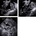
Abnormal nuchal translucency and distinguishing the amnion from the nuchal translucency. A: The fetus is lying supine, and there is a linear structure (arrow) behind the fetal neck that might represent an abnormal nuchal translucency (calipers). On this image, it is unclear whether this linear structure represents the amnion or the skin surface. B: Shortly thereafter, the fetus jumped up, separating the skin surface (arrowhead) from the amnion (arrow), confirming that the nuchal translucency (calipers) is abnormally thick.
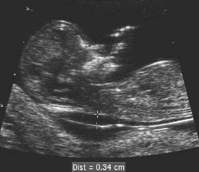
Figure 7.1.4
Abnormal nuchal translucency. Sagittal image of fetus showing abnormally thickened nuchal translucency (calipers), measuring 3.4 mm.
7.2. Thickened Nuchal Fold (16–20 Weeks of Gestation)
Description and Clinical Features
Neonates with trisomy 21 (Down syndrome) often have thickening of the soft tissues in the posterior neck. This has a prenatal analog because thickening of these soft tissues (the nuchal fold) at approximately 16–20 weeks of gestation has been found to be associated with a 10–25-fold increased risk of trisomy 21. When a thickened nuchal fold is found in a 16–20-week fetus, the parents should be counseled about the risk of aneuploidy and offered karyotype testing via amniocentesis.
Sonography
The nuchal fold should be routinely measured on sonograms performed between 16 and 20 weeks of gestation. It is measured on a transverse view of the fetal head in a plane that is close to axial but angled slightly coronally to include the cerebellum and occipital bone. The measurement is taken from just outside the occipital bone to the outer skin surface. A nuchal fold measurement of 6 mm or larger is abnormal (Figure 7.2.1) and should prompt a careful sonographic search for other anomalies, especially those associated with trisomy 21.
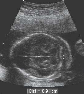
Figure 7.2.1
Thickened nuchal fold. Angled axial view through the fetal head, taken in a plane that includes the cerebellum and occipital bone, demonstrates an abnormally thickened nuchal fold (calipers), measuring 9.1 mm in thickness.
7.3. Cystic Hygroma
Description and Clinical Features
A cystic hygroma is a uni- or multilocular subcutaneous mass filled with lymphatic fluid. It most commonly occurs in the posterior neck, although it can be seen elsewhere in the body. A cystic hygroma results from a localized area of lymphatic dysplasia, with dilation of, or leakage from, lymphatic vessels. Sometimes, the subcutaneous lymphatic dysplasia is widespread, with fluid collections and/or subcutaneous edema encompassing the entire fetal body (generalized cystic hygroma or lymphangiectasia). Even when generalized, the largest fluid collections are typically in the posterior neck.
Fetuses with Turner syndrome (45X karyotype), trisomy 21 (Down syndrome), trisomy 13, and trisomy 18 are at elevated risk of cystic hygromas or generalized lymphangiectasia, particularly during the latter part of the first trimester and the beginning of the second trimester. In view of this, the sonographic finding of cystic hygroma should prompt genetic counseling. Regardless of the fetal karyotype, fetuses with severe generalized cystic hygroma have high mortality rates.
Sonography
A cystic hygroma appears sonographically as a cystic mass in the subcutaneous tissues, generally in the posterior neck, often with septations (Figure 7.3.1). Cystic hygromas can also occur elsewhere in the body (Figures 7.3.2, 7.3.3, and 7.3.4).

Figure 7.3.1
Posterior neck cystic hygroma with septations at 12 weeks of gestation. A: Sagittal image of fetus showing marked expansion of the tissues behind the neck with fluid (calipers). B: Transverse image of neck showing a subcutaneous cystic mass (arrows) that contains septations (arrowheads).
Related posts:


Stay updated, free articles. Join our Telegram channel

Full access? Get Clinical Tree


