Name
Contents
Anatomic location
Supraorbital foramen
Supraorbital nerve (CN V 1 ) and vessels
Frontal bone – superior to the orbit and inferior to the superciliary arch
Superior orbital fissure
CN III, CN IV, CN V 1 , CN VI, ophthalmic vein, and sympathetic fibers from the cavernous plexus
Between lesser and greater wings of the sphenoid bone
Optic canal
Optic nerve (CN II) and ophthalmic artery
Superior aspect of the body and small wing of the sphenoid bone
Cavernous sinus
CN III, CN IV, CN V 1 , CN V 2 , CN VI, internal carotid artery, and sympathetic plexus
Lateral to the sella turcica, on superior portion of the body of the sphenoid bone, and anterior to apex of the petrous part of the temporal bone
Foramen rotundum
Maxillary nerve (V 2 )
Within the anteromedial portion of the sphenoid bone and inferolateral to the superior orbital fissure
Inferior orbital fissure
Zygomatic branch of V 2 and ascending branches of pterygopalatine ganglion
Junction of sphenoid bone and maxilla in the floor of the orbit
Internal auditory canal
CN VII, CN VIII, and nervous intermedius (VII)
Opening in the posterior portion of the petrous temporal bone
Meckel’s cavea
Trigeminal ganglion (CN V)
Bounded by clivus medially, lateral wall of the cavernous sinus superomedially, cerebellar tentorium superolaterally, and apex of the petrous temporal bone inferolaterally
Pterygopalatine fossa (PPF)
Maxillary nerve (V 2 ), Vidian nerve (junction of greater and deep petrosal nerves), terminal portion of the maxillary artery, and pterygopalatine ganglion
Bounded by the infratemporal surface of the maxilla anteriorly, root of the pterygoid process posteriorly, palatine bone medially, pterygomaxillary fissure laterally, and pyramidal process of the palatine bone inferiorly
Palatovaginal canal
Pharyngeal branch of V 2
Connects sphenoid and palatine bones connecting nasopharynx to the pterygopalatine fossa
Zygomaticofacial foramen
Zygomaticofacial nerve (V 2 )
Malar/lateral surface of the zygomatic bone
Foramen ovale
Mandibular nerve (V 3), lesser petrosal nerve (branch of IX), accessory meningeal artery, emissary veins from the cavernous sinus, and the otic ganglion
Posterior portion of the greater wing of the sphenoid bone, posterolateral to foramen rotundum, and anteromedial to foramen spinosum
Sphenopalatine foramen
Nasopalatine nerve (V 2 ) and sphenopalatine vessels
Processes of the superior border of the palatine bone and superiorly by the body of the sphenoid bone
Pterygoid/vidian canal
Nerve (junction of greater and deep petrosal nerves), artery, and vein of the pterygoid canal
Within medial pterygoid plate of the sphenoid bone
Foramen spinosum
Meningeal branch of V 3 and middle meningeal vessels
Posteromedial to the foramen ovale, medial to the mandibular fossa, and anterior to the Eustachian tube
Greater petrosal foramen
Greater petrosal nerve (VII) and petrosal branch of middle meningeal artery
An oblique extension of the facial canal emerging from the anterior surface of the petrous temporal bone
Tympanic canaliculus
Tympanic nerve or nerve of Jacobson (IX)
Small canal within the ridge of the temporal bone separating the carotid canal from the jugular foramen
Carotid canal
Internal carotid artery and carotid plexus of nerves
Within the temporal bone, anterolateral to the jugular foramen, and mediolateral to the Eustachian tube
Jugular foramen
Pars Nervosa – CN IX, tympanic nerve or nerve of Jacobson (IX), and inferior petrosal sinus
Posterior to the carotid canal at the intersection of the petrous temporal and occipital bones. Pars nervosa is the anteromedial portion, while the pars vascularis is the larger posteromedial portion
Pars vascularis – CN X, CN XI, auricular branch of X or Arnold’s nerve, superior bulb of the internal jugular vein, and meningeal branches of ascending pharyngeal and occipital arteries
Greater palatine foramen
Greater palatine nerve (V 2 ) and descending palatine vessels
Foramen at the termination of the greater palatine canal which begins in the inferior PPF passing through the sphenoid and palatine bones to the posterior angle of the hard palate
Infraorbital foramen
Infraorbital nerve (V 2 ) and vessels
Opening in the maxillary bone inferior to the infraorbital margin of the orbit
Hypoglossal canal
CN XII
Superior to the foramen magnum and between the basiocciput and jugular process of the occipital bone
Lesser palatine foramen
Lesser palatine nerve (V 2 )
Posterior to the greater palatine foramen and within the pyramidal process of the palatine bone
Mandibular foramen
Inferior alveolar nerve (V 3 ) and artery
Foramen on internal surface of the ramus of the mandible
Mental foramen
Mental nerve (V 3 ) and vessels
Anterior surface of the mandible
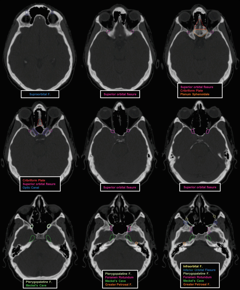
Fig. 1
Important foramina, fossa, and canals of the cranium and skull base
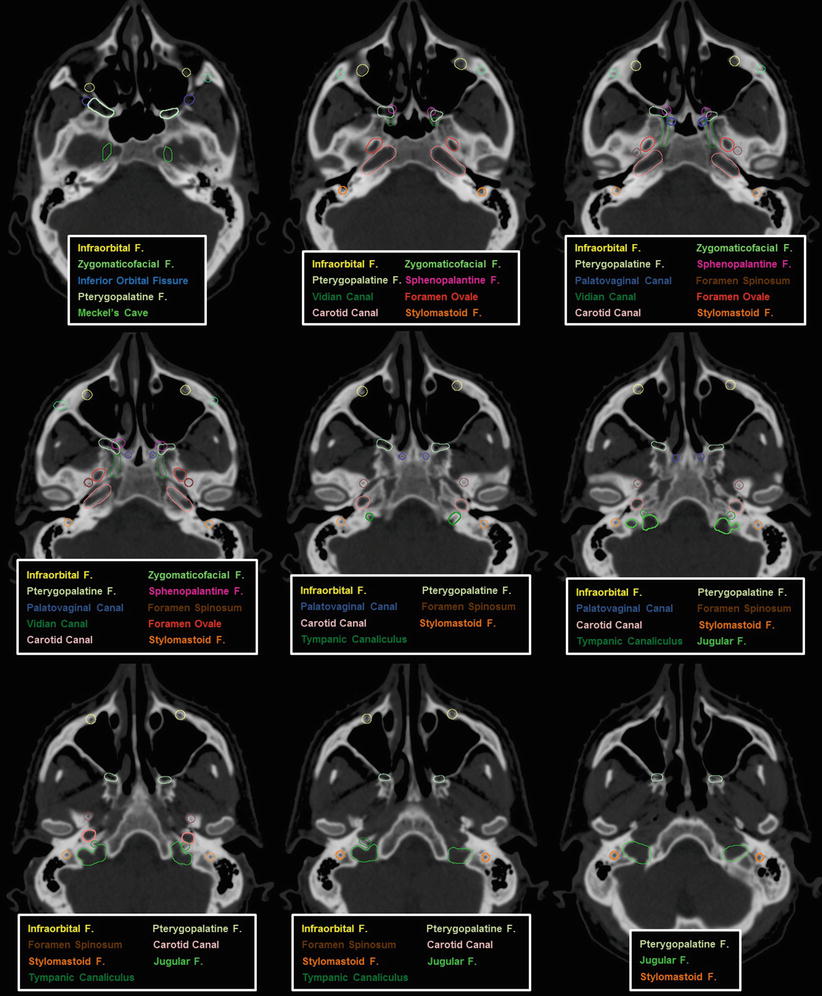
Fig. 2
Important foramina, fossa, and canals of the cranium and skull base
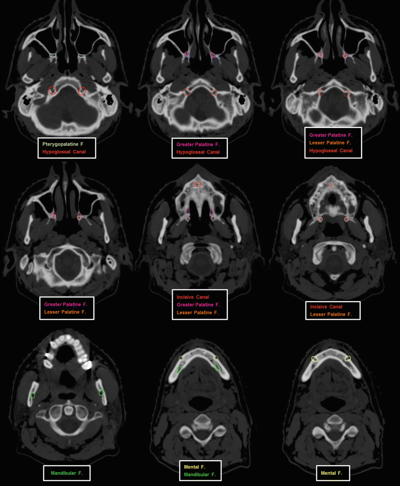
Fig. 3
Important foramina, fossa, and canals of the cranium and skull base
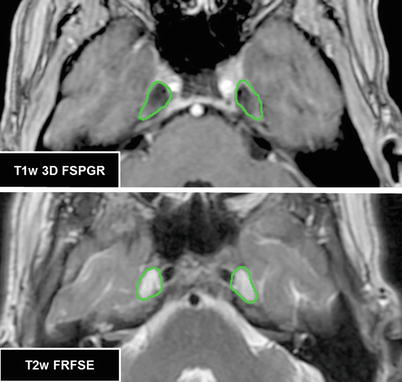
Fig. 4
Meckel’s cave which contains the trigeminal ganglion is easily identifiable on a number of MRI sequences. In this figure, Meckel’s cave (in green) is seen as a hypointense structure on a T1-weighted FSPGR image and a hyperintense fluid-filled structure on a T2-weighted fast relaxation fast spin echo (FRFSE) image
2.2 The Cranial Nerve Pathways
Cranial Nerve I: The cribriform plate of the ethmoid bone is the primary landmark to identify the location of CN I. It forms the roof of the nasal cavities. Small foramina in the cribriform plate allow passage of numerous olfactory nerves from the roof of the nasal cavity, superior portion of the nasal septum, and superior nasal concha. After passing through the cribriform plate, these nerves traverse the dura mater and subarachnoid space before ending in the olfactory bulb, which is situated under the basal surface of the frontal lobe.
 Identification of the olfactory nerve is not routinely performed in planning for radiotherapy. It may be important in the treatment of certain skull base tumors such as olfactory groove meningiomas and esthesioneuroblastomas as recently reported by the University of Pittsburgh Medical Center (Gande et al. 2014). The cribriform plate is seen in Fig. 1.
Cranial Nerve II: The axons of the retinal ganglion cells in the eye are the basis of CN II which originates where they pass through the sclera. The nerve passes posteromedially beneath the superior rectus muscle and between the medial and lateral rectus muscles. It exits the orbit through the optic canal located in the sphenoid bone. Intracranially, the nerve passes superomedially to the ophthalmic artery and terminal segments of the internal carotid artery. The nerve ends at the optic chiasm which lies superior to the sella turcica, anterior to the pituitary stalk, and inferior to the supra-optic recess of the third ventricle. The central retinal artery and vein travel with the optic nerve within the meninges until they rejoin the ophthalmic artery and superior ophthalmic vein, respectively.
 The optic nerve is not often a treatment target unless it is grossly involved with disease (see Case 6). More often it is contoured as an organ at risk (OAR) for treatment planning assessment. Use of a fused T1-weighted MR image is best for delineating the chiasm. Figure 5 illustrates the path of CN II with respect to the structures discussed above.
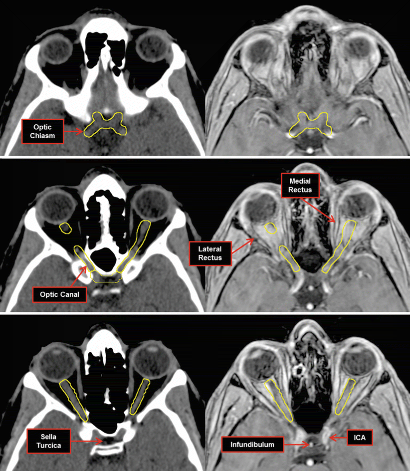
Fig. 5
Path of the optic nerve (yellow) into the optic chiasm. Images on the left are non-contrast CT scans with corresponding images from a T1w 3D FSPGR MRI on the right. ICA internal carotid artery
Cranial Nerve III: The oculomotor nerve originates from two nuclei (somatic and visceral motor) located in the midbrain at the level of the superior colliculus and anterior to the cerebral aqueduct. The nerve exits the midbrain into the interpeduncular fossa medial to the basilar artery. It immediately passes between the posterior cerebral artery and superior cerebellar artery. The nerve enters the cavernous sinus lateral to the internal carotid artery and superior to CN IV. It enters the orbit through the superior orbital fissure wherein it divides into the superior and inferior divisions to supply motor innervation to its target extraocular muscles. The inferior division carries the parasympathetic fibers to the ciliary and constrictor pupillae muscles.
 The oculomotor nerve is typically not contoured independent of other structures. If there is concern for involvement of the cavernous sinus, all structures within are at risk and the whole region is covered by a treatment volume. As shown in Fig. 6, the path of CN III is traceable into the orbit.
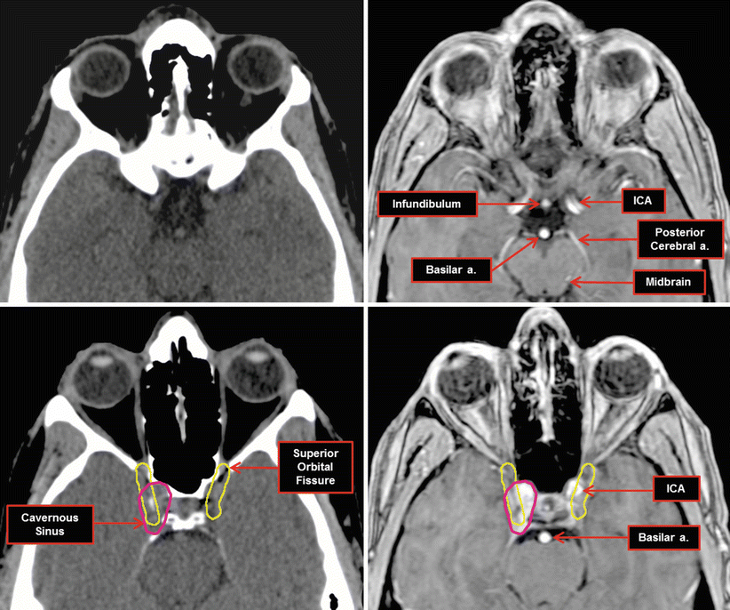
Fig. 6
Path of cranial nerve III, the oculomotor nerve (yellow), from the midbrain through the cavernous sinus and finally into the orbit through the superior orbital fissure. Images on the left are non-contrast CT scans with corresponding images from a T1w 3D FSPGR MRI on the right. ICA internal carotid artery
Cranial Nerve IV: The trochlear nerve motor nucleus is in the periaqueductal gray matter of the midbrain at the level of the inferior colliculus. The nerve fibers emerge dorsally from the midbrain, travelling anterolaterally between the posterior cerebral artery and superior cerebellar arteries. It enters the lateral wall of the cavernous sinus inferolateral to CN III. To enter the orbit, the nerve passes medially through the superior orbital fissure below the anterior clinoid process and CN III.
 Similar to CN III, the trochlear nerve is not contoured independent of other structures. If there is clinical or radiographic involvement of the nerve, the contents of the entire cavernous sinus are often involved and treated. Figure 7 depicts the course approximate of CN IV into the orbit.
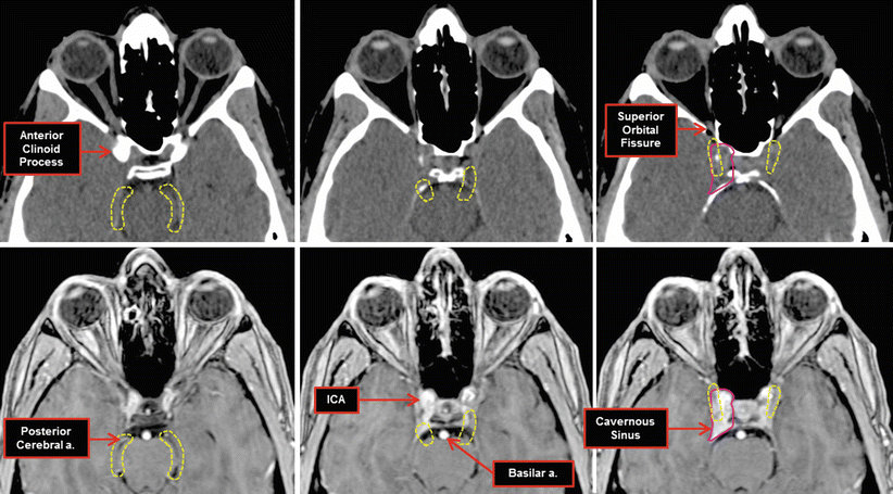
Fig. 7
Approximate path of cranial nerve IV, the trochlear nerve (dashed yellow), from the dorsal midbrain through the cavernous sinus and finally into the orbit through the superior orbital fissure inferior to the anterior clinoid process. Images on top are non-contrast CT scans with corresponding images from a T1W 3D FSPGR MRI on the bottom. ICA internal carotid artery
Cranial Nerve V: The trigeminal nerve originates from 4 nuclei – 1 motor and 3 sensory. The locations and paths of these nuclei are beyond the scope of this chapter and can be reviewed elsewhere (Bathla and Hegde 2013; Moore and Dalley 1999). CN V emerges from the pons as essentially two separate nerves: a small motor and large sensory root. The collection of cranial nerve roots enters Meckel’s cave (Fig. 8) wherein the sensory branches coalesce into the trigeminal ganglion, while the motor root travels medially and rejoins the sensory portion of V3 to create the mandibular nerve. The trigeminal ganglion splits into the 3 branches of CN V: the ophthalmic nerve (V1), maxillary nerve (V2), and the sensory component of the mandibular nerve (V3). Both V1 and V2 pass through the lateral wall of the cavernous sinus inferolateral to CN IV and III.
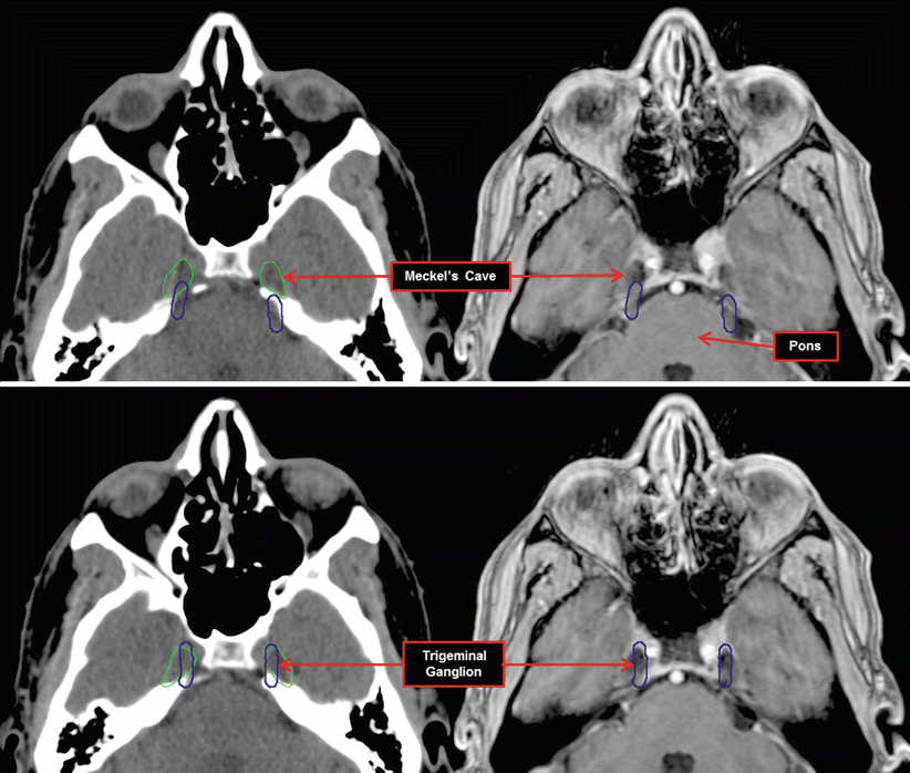

Fig. 8
The trigeminal trunk is illustrated emerging from the pons (blue) and entering Meckel’s cave where it forms the trigeminal ganglion. Images on the left are non-contrast CT scans with corresponding images from a T1w 3D FSPGR MRI on the right
The ophthalmic nerve (V 1 ) enters the orbit through the superior orbital fissure where it divides into its terminal branches: the lacrimal, frontal, and nasociliary nerves. The lacrimal nerve follows the course of the lateral rectus along its superior edge until it reaches the lacrimal gland. The frontal nerve passes medial to the lateral rectus muscle and travels superior to the superior rectus finally terminating as the supratrochlear and supraorbital nerves. The nasociliary nerve travels medially in the orbit until it divides into its terminal branches that supply sensation to nasal cavity and paranasal sinuses.
 If there is perineural spread along V1, the clinical treatment volume (CTV) encompasses nearly the whole of the orbit to Meckel’s cave. This covers both retrograde and anterograde spread along the terminal branches. As seen in Fig. 9, the CTV for the lacrimal nerve includes the lateral rectus and extends superiorly eventually encompassing the lacrimal gland on more superior axial slices. A CTV for the frontal nerve includes the ophthalmic artery and superior rectus. It also encompasses the supraorbital foramen in the most superior aspect. The nasociliary nerve spreads out into multiple branches along the medial wall of the orbit of which a CTV would include.
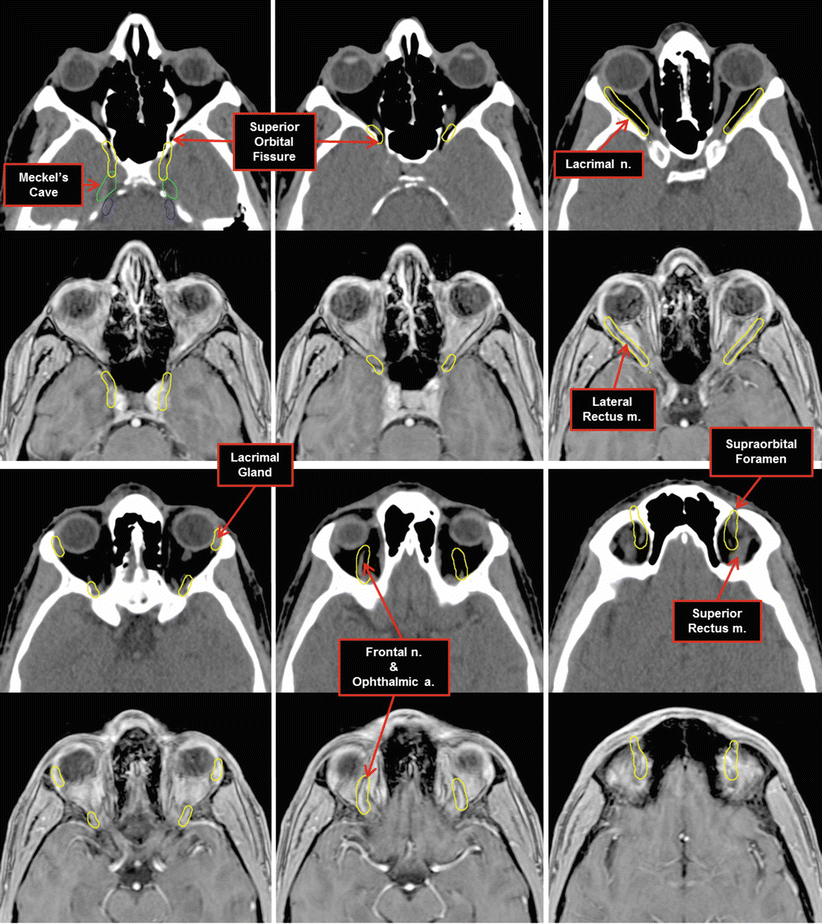
Fig. 9
The approximate pathway and branches of the ophthalmic branch (CN V1) of the trigeminal nerve (CN V) are shown here after exiting Meckel’s cave. Images on the top are non-contrast CT scans with corresponding images from a T1w 3D FSPGR MRI on the bottom. CN V1 yellow
After traversing the cavernous sinus, the maxillary nerve (V 2 ) exits through the foramen rotundum and enters the pterygopalatine fossa. Prior to entering the foramen rotundum, V2 gives off the middle meningeal nerve which enters the foramen spinosum. Within the pterygopalatine fossa, V2 divides into its terminal branches: zygomatic, greater palatine, lesser palatine, nasopalatine, posterior-superior alveolar, and infraorbital nerves. The zygomatic nerve enters the orbit through the inferior orbital fissure and supplies sensation to the skin over the zygomatic and temporal bones. Sphenopalatine branches of V2 directly supply the pterygopalatine ganglion and continue into the greater and lesser palatine nerves. The greater palatine nerve moves inferiorly from the pterygopalatine fossa and enters the hard palate through the greater palatine foramen. The lesser palatine nerve follows a similar course and emerges to innervate the soft palate, tonsils, and uvula through the lesser palatine foramen. The posterior-superior alveolar nerve arises from V2 prior to entering the infraorbital groove and descends to supply a portion of the gums and teeth. The infraorbital nerve enters the orbit through the inferior orbital fissure and enters the infraorbital canal exiting to innervate the lower eyelid, upper lip, and part of the nasal vestibule through the infraorbital foramen.
 The CTV for CV V2 is illustrated in Fig. 10. Here the CTV includes all the major branches of V2 except for the nasopalatine and posterior-superior alveolar nerve. Inclusion of these branches is important when there is gross tumor involvement of the pterygopalatine fossa (see Case 6). A CTV that includes the nasopalatine nerve would encompass the nasal septum down to the incisive foramen in the anterior hard palate. The osseous structures around the nerves are included in the CTV given variability in nerve position and difficult visualization on MRI unless there is perineural spread.
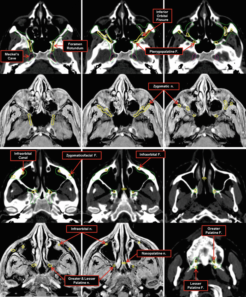
Fig. 10
The pathway and branches of the maxillary branch (CN V2) of the trigeminal nerve (CN V) are shown here after exiting Meckel’s cave. The CTV is shown in green. Images on the top are non-contrast CT scans with corresponding images from a T1w 3D FSPGR MRI on the bottom. CN V2 yellow
The mandibular nerve (V 3 ) exits Meckel’s cave inferiorly through the foramen ovale. Upon exiting the foramen ovale, the nerve splits into an anterior and posterior branch. The anterior nerve supplies the masseteric, deep temporal, buccal, and medial and lateral pterygoid nerves. The posterior division divides into the auriculotemporal, lingual, and inferior alveolar nerves and motor branches to the mylohyoid and anterior belly of the digastric muscles. The inferior alveolar nerve enters the mandibular foramen and supplies sensation to the mandibular teeth and finally the lower lip and chin after it exits the mandible through the mental foramen as the mental nerve.
 The CTV for CN V3 is defined by the parapharyngeal spaces containing the vessels and best highlighted by a T2-weighted MRI (Fig. 11). The CTV begins superiorly at Meckel’s cave and includes the foramen ovale and these parapharyngeal spaces to the mandibular foramen and the sublingual space along the lingual artery. If there is gross perineural spread, the muscles innervated by the V3 should be included in the CTV (Cases 2 and 5).
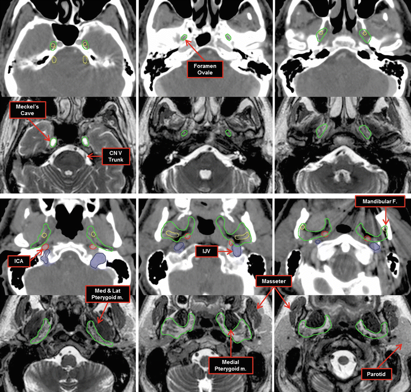
Fig. 11
The pathway of the mandibular nerve, CN V3 (yellow), of the trigeminal nerve (CN V) is shown here after exiting Meckel’s cave and entering the foramen ovale. The CTV is indicated in green. Images on the top are contrast CT scans with corresponding images from a T2w 3D MRI on the bottom. ICA internal carotid artery, IJV internal jugular vein
Cranial Nerve VI: The abducens nerve arises in the medial aspect of the pons near the floor of the 4th ventricle and adjacent to the motor nucleus of CN VII. It emerges into the pontine cistern at the pontomedullary junction. It travels anteriorly along the basilar artery and enters the cavernous sinus through Dorello’s canal (Fig. 12) after bending over the petrous portion of the temporal bone. In the cavernous sinus it is immediately posterolateral to the internal carotid artery and medial to CN III and IV. It enters the orbit through the medial aspect of the superior orbital fissure where it innervates the lateral rectus muscle. The approximate pathway through the cavernous sinus is seen in Fig. 13.
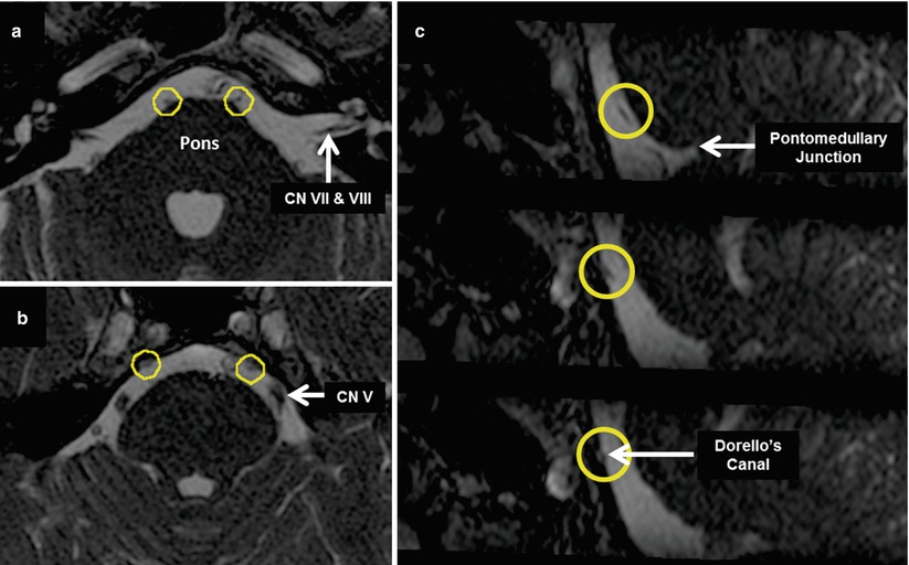
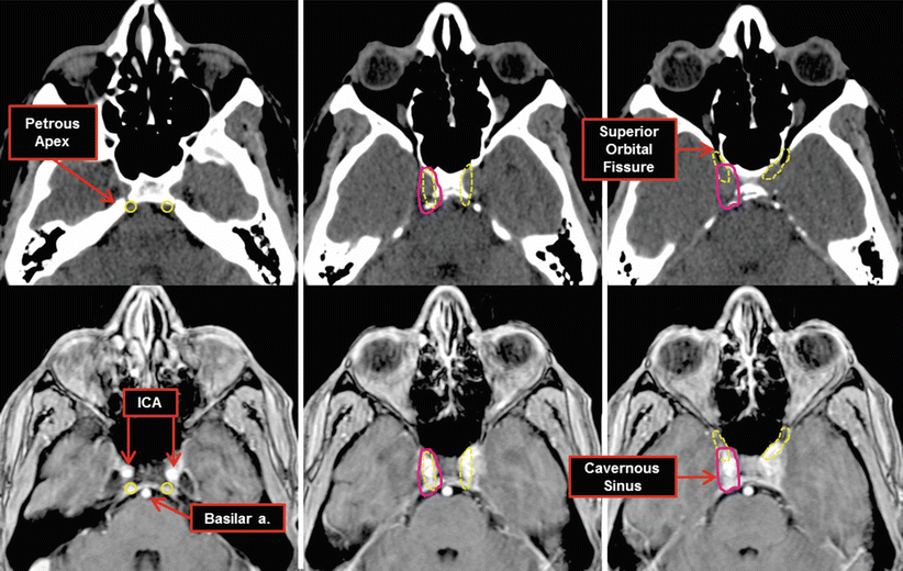

Fig. 12
The abducens nerve (CN VI) as it exits the pontomedullary junction as seen on a 3D FIESTA MRI (a). (b, c) show CN VI entering Dorello’s canal after traveling superiorly in the pontine cistern. Additional cranial nerves are also seen in these images. Yellow circles CN VI

Fig. 13
The approximate pathway of the abducens nerve, CN VI (dashed yellow), is shown here after entering Dorello’s canal and crossing the petrous apex. Images on the top are non-contrast CT scans with corresponding images from a T1w 3D FSPGR MRI on the bottom. ICA internal carotid artery
 The abducens nerve is difficult to image except on very dedicated scans as shown in Fig. 12. If it is clinically grossly involved with disease, the entire cavernous sinus is included in the CTV.
Cranial Nerve VII: The facial nerve originates from two separate nerves exiting the brainstem at the cerebellopontine angle. The motor component arises from the motor nucleus in the dorsal pons. The sensory component, often called the nervus intermedius, arises from a combination of inputs from the spinal nucleus of CN V, the superior salivary nucleus, and the nucleus solitarius. The two nerves cross the cerebellopontine cistern and join at the internal acoustic meatus. The facial nerve travels with CN VIII until it enters the facial/fallopian canal and travels anterolaterally. At the geniculate ganglion the nerve abruptly changes direction forming the first genu. Here, the greater petrosal nerve branches off the facial nerve and travels through the greater petrosal foramen into the middle cranial fossa until it reaches the vidian canal where it forms the vidian nerve and finally reaches the pterygopalatine ganglion. The facial nerve travels posteriorly along the medial wall of the tympanic cavity wherein it gives rise to the nerve to the stapedius. At the end of the tympanic cavity is the second genu where the nerve turns 90° inferiorly and enters the mastoid bone. In this segment, the facial nerve gives rise to the chorda tympani which eventually joins the lingual nerve of CN V 3 to supply taste to the anterior two-thirds of the tongue. The facial nerve then exits the cranium through the stylomastoid foramen and enters the substance of the parotid gland. The nerve terminates in six branches: the posterior auricular, temporal, zygomatic, buccal, mandibular, and cervical nerves. Additional information on the anatomy and path of the facial nerve can be found elsewhere (Gilchrist 2009; Veillona et al. 2010).
Get Clinical Tree app for offline access

 If there is facial nerve involvement of disease, the CTV typically includes the entire parotid gland as the 6 terminal branches are within its substance. The CTV continues superiorly through the stylomastoid foramen in the mastoid bone. Inclusion of the chorda tympani in the CTV may also be important based on clinical considerations. Unless there is gross perineural invasion of CN VII into the stylomastoid foramen, the CTV can stop below the 2nd genu given additional toxicity to the cochlea and inner ear. Case 1 demonstrates a CTV for CN VII involvement. The course of the nerve within the mastoid bone is illustrated in Fig. 14.
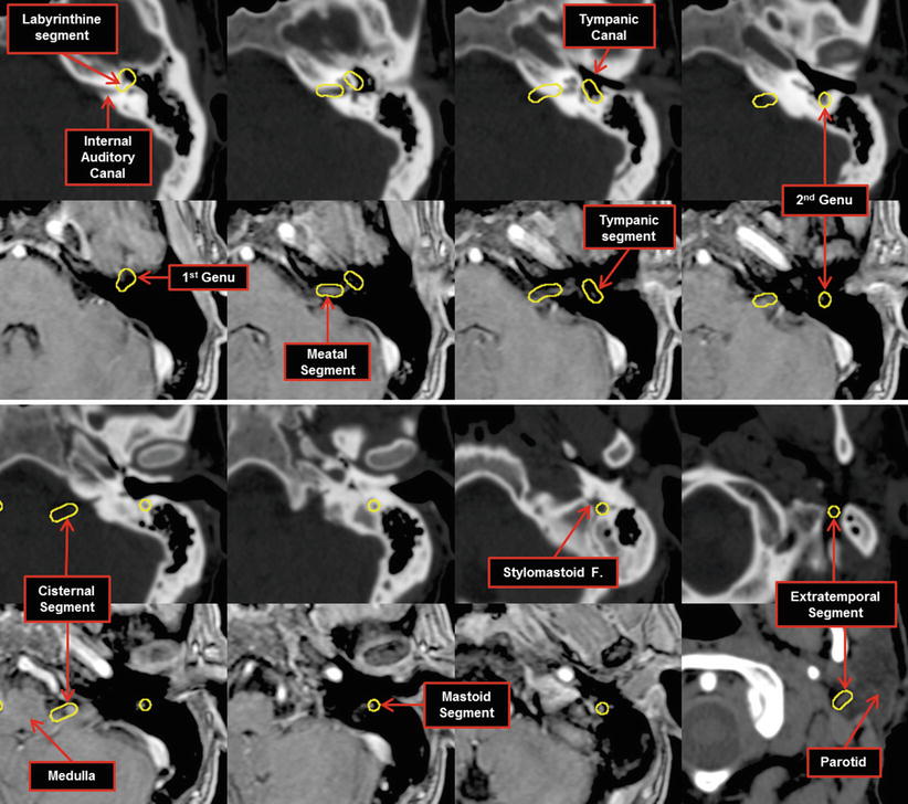
Fig. 14
Axial slices through the mastoid bone with the pathway of the facial nerve (CN VII) identified. Images on the top are non-contrast CT scans with corresponding images from a T1w 3D FSPGR MRI on the bottom
Stay updated, free articles. Join our Telegram channel

Full access? Get Clinical Tree


