(1)
Memorial Sloan Kettering Cancer Center, New York, 10065, NY, USA
1 Anatomy
The paranasal sinuses are composed of seven bones (ethmoid, maxilla, palatine, lacrimal, pterygoid plate of sphenoid, nasal, and inferior turbinate), four paired sinuses (frontal, ethmoid, maxillary, and sphenoid), and complex networks of nervous, vascular, and lymphatic structures.
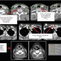
Fig. 1
Representative normal anatomy from axial CT slices
2 Patterns of Spread
Paranasal sinus cancers often spread via local extension into adjacent sinuses and bony structures. The paranasal sinuses (and nasal cavity) are all interconnected via multiple ostia and are separated by thin septi allowing for invasion into adjacent air cavities.
Maxillary Sinus (as a representative example):
Medial infrastructure lesions invade the nasal cavity early via the porous medial wall.
Lateral infrastructure lesions erode the lateral wall of the antrum and may present as a submucosal mass in the maxillary gingiva.
Posterior infrastructure lesions may invade the infratemporal fossa or extend into the pterygopalatine fossa and pterygoid plates. These lesions may invade the orbit by direct superior extension or via extension into the ethmoids.
Suprastructure lesions spread either laterally, invading the malar process of the maxilla and the zygoma, or medially, invading the nasal cavity and ethmoid sinuses.
Involvement of nerve branches (see Table 1) by tumor often leads to numbness and paresthesias in the skin and mucous membranes of this region. It is of critical importance if cranial nerve involvement is present to cover the involved nerve(s) back to the skull base.
Table 1
Anatomy of paranasal sinuses
Sinus
Overview and boundaries
Blood supply and innervation
Frontal
Located in the frontal bone superior to the orbits in the forehead
Supraorbital and supratrochlear arteries of the ophthalmic artery
Funnel-shaped structure
Supraorbital and supratrochlear nerves of the first division of the trigeminal nerve
An upward extension of anterior ethmoid cells
The posterior wall separates the sinus from the anterior cranial fossa. The floor is formed from the upper part of the orbit
Sphenoid
Located in the sphenoid bone at the center of the head
Sphenopalatine artery (and branches), except for the planum sphenoidale, which is supplied by the posterior ethmoidal artery
Closely related to many critical structures: pituitary gland, superiorly; optic nerve and carotid artery, laterally; and nerve(s) of the pterygoid canal at the floor
Branches of the first and second divisions of the trigeminal nerve
May extend as far as the foramen magnum
Maxillary
Pyramidal shaped (the base is along the nasal wall and the apex points laterally toward the zygoma)
Branches of the internal maxillary artery (infraorbital, alveolar, greater palatine, and sphenopalatine arteries)
The roof is the floor of the orbit. The floor is made from the alveolar process of the maxilla and hard palate
Branches of the second division of the trigeminal nerve, the infraorbital nerve, and the greater palatine nerves
Behind the posteromedial wall of this sinus lays the pterygopalatine fossa (houses important neurovascular structures and communicates with skull base foramina)
The infratemporal fossa lies behind the posterolateral wall of the maxillary sinus
Ethmoid
Located in the ethmoid bone, forming multiple air cells (~6–15) between the eyes
Anterior and posterior ethmoidal arteries from the ophthalmic artery (internal carotid system), as well as by the sphenopalatine artery from the terminal branches of the internal maxillary artery (external carotid system)
The ethmoid cells are shaped like pyramids and are divided by thin septa
V1 supplies the more superior aspect with V2 innervating the inferior regions. Parasympathetic innervation is via the Vidian nerveRelated posts:

Stay updated, free articles. Join our Telegram channel

Full access? Get Clinical Tree

 Get Clinical Tree app for offline access