Fig. 1
Primary lymphatic sites of bladder carcinoma: Distribution of positive nodes after cystectomy and PLND in 60 patients. The noted areas are the external iliac (Ia) distal and (Ib) proximal, obturator fossa (IIa) distal and (IIb) proximal, internal iliac (IIIa) distal and (IIIb) proximal, common iliac (IVa) distal and (IVb) proximal, (V) para-aortic/cava (Roth et al. 2010)
In an analysis of the primary lymphatic sites of bladder carcinoma, it was discovered that 92 % of positive lymph nodes were distal and caudal to where the ureter crosses the common iliac arteries (Roth et al. 2010). All of those that were positive proximal to the crossing of the ureter over the common iliac arteries also showed endopelvic positive lymph nodes on extended pelvic lymph node dissection (PLND). Therefore, coverage superior to the ureters crossing the common iliac arteries would not aid in local control.
2 Diagnostic Workup Relevant for Target Volume Delineation
Painless hematuria and voiding symptoms often initiate a workup which includes cystoscopy with cytology and urinalysis revealing carcinoma. Initial workup is recommended to include digital rectal and bimanual examinations as well as imaging of the upper tract. The presence of ureteral obstruction, hydronephrosis, or a nonfunctioning kidney predicts muscle-invasive disease in 90 % of cases (Messer et al. 2011; Hatch and Berry 1986). The importance of a proper workup cannot be overstated for candidates for bladder preservation therapy since they are commonly understaged compared to those that undergo surgery.
For patients who may be candidates for bladder preservation, a workup should include cystoscopy with a detailed description or illustrative mapping which provides direct visualization and enumeration of any biopsy-positive disease. Descriptors including size and location of any tumors are vital for target delineation.
Patients with Ta or low-grade T1 tumors do not require an extensive metastatic workup. Those with muscle-invading tumors, however, have a risk of occult metastatic disease of up to 50 % and should undergo chest imaging and bone scanning if alkaline phosphatase is elevated.
For patients with muscle-invasive disease, imaging of the upper tract is recommended and can include more common studies such as intravenous pyelogram or CT urography but may also include MRI urogram, ureteroscopy, or renal ultrasound with retrograde pyelogram. Locally advanced disease imaging with CT or MRI is imperative for proper staging, and MRI has shown improved specificity over CT scan due to its high soft tissue contrast and also has the advantage that contrast agents are non-nephrotoxic (Zhang et al. 2007). The contrast between fat and tissue is best defined on T1-weighted imaging or T1 subtraction sequences. Figure 2a shows how MRI can be used for staging as well as target delineation of the gross tumor volume (GTV) for a patient treated with IMRT. Notably, the contrast between the fat and tissue may be lost on the normal bladder wall several months after radiation treatment and limits the usefulness of follow-up MRI scans.


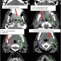
Fig. 2
(a) MRI for patient showing axial T1-post-contrast subtraction sequence demonstrating invasion beyond the wall of bladder (red arrow) and loss of fat plane (white arrow). (b) CT contours for patient in Fig. 2a using IMRT. Target delineation CTV364.5 Gy (red), PTV1 51 Gy (blue), and PTV254 Gy (orange) in a patient with muscle-invasive bladder cancer with focus at the primary site. GTV based on MRI and CT imaging. (c) CT contours for patient in Fig. X-X using IMRT. Target delineation CTV364.5 Gy (red), PTV1 51 Gy (blue), and PTV254 Gy (orange) in a patient with muscle-invasive bladder cancer with focus at the primary site. Please note that these are representative slices and not all slices are included. Please note normal structure contours include bladder (yellow), seminal vesicles (green), and rectum (brown)
3 Simulation and Daily Localization
CT simulation using 3 mm or less slice thickness should be performed prior to planning. For 3DCRT, set up the patient in supine position with the bladder emptied just prior to simulation for reproducibility. Similarly, patients are asked to void the contents of the rectum for daily reproducibility. When using 3DCRT technique, a sequential boost is added to the initial clinical target volume (CTV). A new simulation may be helpful with a partially distended bladder to limit dose to the entire bladder as well as the rectum depending on the location of the tumor. Many institutions continue the boost with the bladder empty since this provides reproducibility.
If using IMRT with image guidance, the simulation is performed with the bladder partially full to allow for dose painting to the GTV daily. Bladder filling should be comfortable and reproducible by the patient. On board imaging capable of assessing bladder filling and volume is critical to treating with a full bladder. Simulation may include leg support for patient comfort which can aid in daily setup. Immobilization devices including Vak-Lok or Alpha Cradle may be used and vary by institution. Similarly, frog-leg positioning may be warranted to reduce skinfolds.
IV contrast may be used to help guide the GTV target and vessels; however, renal function must be assessed and fusion of imaging from the patient’s workup may be adequate. A simulation scan from the upper lumbar spine to mid-femur is recommended.
Image registration and fusion applications with previously performed MRI and PET are recommended to help in the delineation of target volumes. However, the bladder mapping in conjunction with the surgeon is imperative for the CTV. Normal tissues should be outlined on all CT slices in which the structures exist. An example of how CT urography may aid in target delineation is provided in Fig. 3.

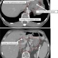
Fig. 3
Target delineation GTV outlined in red is displayed in CT urogram. PTV1 (purple) for lymph nodes is shown prior to expansion for planning. Please note that these are representative slices and not all slices are included. Please note normal structure contours include bladder (yellow), seminal vesicles (green), prostate (pink) and rectum (brown)
Image-guided radiation therapy allows for adjustments to be made based on soft tissue anatomy prior to treatment delivery. Cone-beam CT can be performed in an effort to decrease uncertainty and therefore decrease treatment margins. It also allows for confirmation of bladder filling. While not the standard, IMRT is an option to reduce bowel dose as well as spare uninvolved bladder during a boost.
4 Target Volume Delineation and Treatment Planning
Bladder preservation therapy is a multidisciplinary effort with concurrent chemotherapy and radiotherapy after maximal transurethral resection of bladder tumor (TURBT). Ideal candidates for bladder preservation include those with clinical T2-3a tumors that are unifocal, node negative, and with no evidence of ureteral obstruction or involvement with the ureteral orifice and good bladder function. Ideal candidates should have a complete resection by TURBT (Shipley et al. 1998; Tester et al. 1993).
To properly plan the boost portion of therapy, bladder mapping from the pretreatment cystoscopy should be used as well as any findings on physical exam or imaging that display extravesicular extension.
Suggested target volumes at the GTV and high-risk CTV are detailed in Table 1 for 3DCRT planning (Shipley et al. 1998).
Table 1
Suggested target volumes for bladder preservation (3DCRT)Related posts:

Stay updated, free articles. Join our Telegram channel

Full access? Get Clinical Tree