R testicular seminoma tends to drain to the vena cava and aortocaval nodes.
L testicular seminoma tends to drain to the L renal vein and para-aortic nodes.
2 Diagnostic Workup Relevant for Target Volume Delineation
A suspicious testicular mass should prompt a complete history and physical exam, serum tumor markers (α-fetoprotein, β-human chorionic gonadotropin, and LDH), chemistry panel, and CXR. Always REMEMBER SPERM BANKING.
After orchiectomy, patients should complete staging with a CT of the abdomen and pelvis (and include CT chest if suspicious nodes on CT A/P) and have repeat of beta-hCG, LDH, and AFP.
At some institutions, a PET/CT will be part of initial workup. This could conceivably help with target delineation.
3 Simulation and Daily Localization
Simulation in the supine position with arms in wing-board and wedge under knees.
One could consider using alpha-cradle to help to enhance immobilization.
A clamshell shield must be added to decrease dose to the remnant testicle.
Move the penis out of the field with mesh.
Tattoos must be placed at the level of the isocenter anteriorly and laterally.
IV contrast can be used to help better identify both the vessels and any gross disease.
If available, a PET/CT simulation may be helpful in stage II cases for delineation of gross nodal disease.
4 Target Volume Delineation and Treatment Planning
Stage I: It is the strong recommendation of the National Comprehensive Cancer Network Treatment (NCCN) that all patients with stage I seminoma undergo post-orchiectomy surveillance. Treatment of these patients risks late morbidity, most notably secondary malignancies. However, if a patient refuses active imaging surveillance, based on results from MRC TE 10 and TE 18, patients with stage I seminoma can receive adjuvant radiotherapy confined to para-aortic lymph nodes to a dose of 20 Gy unless there is prior inguinal or scrotal violation (lymphatic alteration) (Fossa et al. 1999; Jones et al. 2005). Fields for stage I disease are outlined below. Besides radiotherapy, carboplatinum ×1 cycle is also an option for stage I patients who refuse surveillance (National Comprehensive Cancer Network 2014).
Stage II: Radiotherapy to a traditional “dog-leg” field is the typical standard of care for stage IIa patients as outlined below; for stage IIb patients, options include radiotherapy as outlined or chemotherapy (etoposide + cisplatinum ×4 cycles or bleomycin, etoposide, cisplatinum ×3 cycles); stage IIc patients should be treated with chemotherapy (same) (National Comprehensive Cancer Network 2014).
Volume recommendations were derived from Wilder et al. Excellent reference (Wilder et al. 2012) (Tables 1 and 2; Figs. 1 and 2).
Table 1
Suggested target volumes for stage IA, IB, or IS
Target volumes
Definition and description
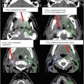
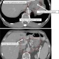
CTV
Based on CT imaging (preferred): contour out the inferior vena cava and aorta from 2 cm below the top of the kidney superiorly to bifurcations inferiorly → provide a 1.2 cm expansion on IVC and 1.9 cm expansion on the aorta → contour out the bone, muscle, and bowel → merge two expanded volumesRelated posts:

Stay updated, free articles. Join our Telegram channel

Full access? Get Clinical Tree

 Get Clinical Tree app for offline access