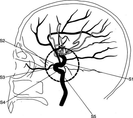
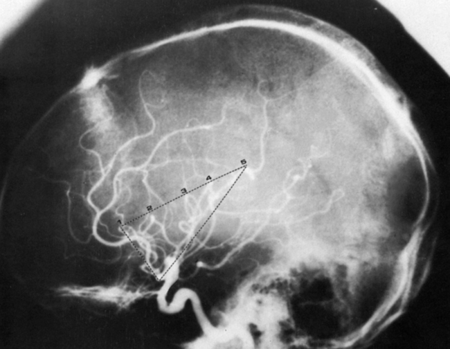
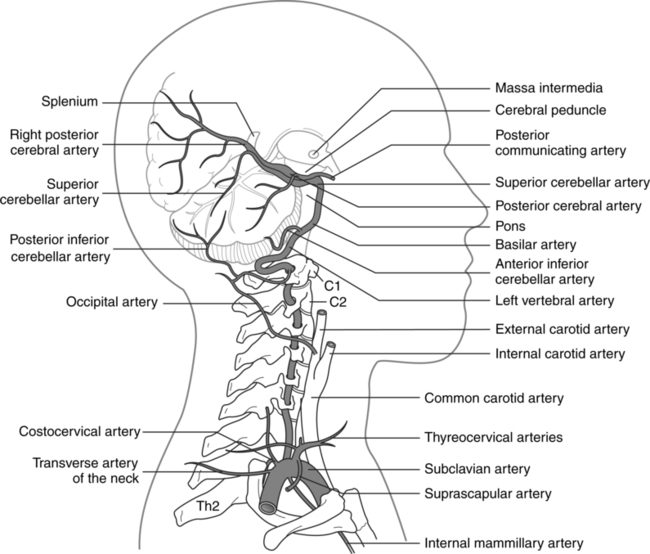
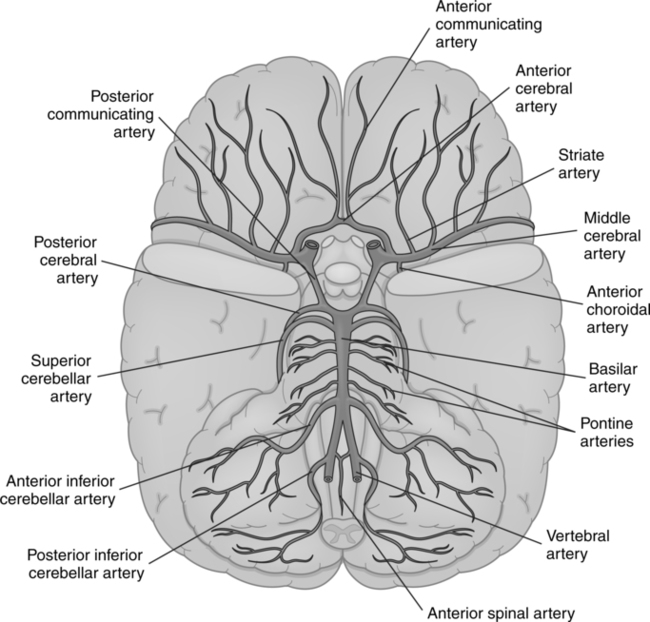
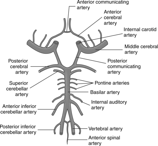
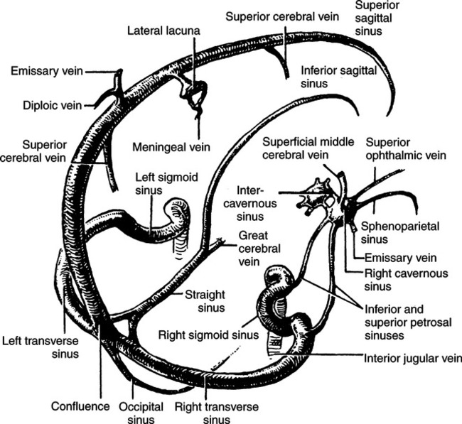
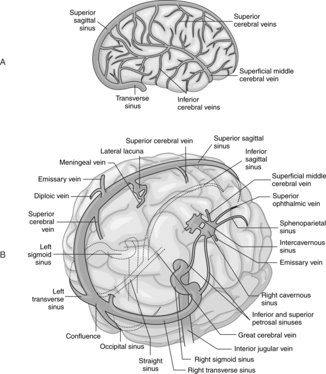
CHAPTER 16 After completing this chapter, the reader will be able to perform the following: A discussion of the cerebrovascular system must begin with the aortic arch, which is where the major vessels originate. In Chapter 15, we saw that the aortic arch has three major branches—the brachiocephalic trunk, the left common carotid artery, and the left subclavian artery (see Fig. 15-1). The brachiocephalic trunk is about 4 to 5 cm long at the upper border of the right sternoclavicular articulation and bifurcates into the right subclavian and right common carotid arteries. The left common carotid artery originates at the highest point of the aortic arch. It ascends to the left sternoclavicular joint, where it enters the neck. The common carotid arteries are unequal in length, with the left being 4 to 5 cm longer than the right. The carotid line, which begins at the sternoclavicular joint and terminates midway between the angle of the mandible and the mastoid process of the temporal bone, defines the course of the carotid artery. At the cranial edge of the thyroid cartilage, the common carotid arteries divide terminally into two main branches—the internal and external carotid arteries. At its bifurcation, there is a dilation of the terminal portion of the common carotid artery and the base of the internal carotid artery; this dilated portion is called the carotid sinus. The carotid sinus is similar to the aortic sinus in that it contains certain receptors that help to control blood pressure. The course of the internal carotid is illustrated in Figure 16-1. The path of the internal carotid artery ascends from the underside of the petrous bone to the peak of the petrous pyramid. This path resembles an inverted “L.” It then curves forward and medially, running laterally to the sphenoidal structures (S5). The artery then enters the cavernous sinus and passes next to the sella turcica (S4). It ascends, passes through the dural roof of the sinus under the base of the anterior clinoid process (S3), and then courses backward, passing under the optic nerve. At this point, the artery lies in the subarachnoid space (S2). It then ascends, dividing into the anterior and middle cerebral arteries (S1). The curving portions of the internal carotid artery, designated S2, S3, and S4, were called the carotid siphon by Moniz. The ophthalmic artery originates at point S3 of the carotid siphon. Two other arteries arise as branches of the carotid siphon—the anterior choroidal and posterior communicating arteries. The isle of Reil is a triangular structure that can be delineated by the middle cerebral artery and its branches, which form the sylvian triangle (Fig. 16-2). This is a very important anatomic landmark; it is usually affected by most mass lesions, and any shift will probably be demonstrated. Another important arterial system that supplies the posteroinferior portion of the brain, brain stem, and cerebellum is the vertebrobasilar system (Fig. 16-3). The vertebral arteries originate as branches of the subclavian arteries. The left is longer than the right, because the right subclavian artery originates as a branch of the brachiocephalic artery. The vertebral arteries run through the cervical region and then pass through the foramen magnum to enter the skull. Running parallel to each other for a short distance, the vertebral arteries begin to converge and then unite to form the basilar artery. The basilar artery begins at the lower border of the pons and terminally divides into the paired posterior cerebral arteries. In general, the basilar artery can be found traveling in the longitudinal groove at the front of the pons (Fig. 16-4). A short distance after the union of the vertebral arteries, the paired anteroinferior cerebellar arteries arise as branches of the basilar artery. There are numerous pontine branches (paramedian arteries) coursing perpendicularly into the pons. As their name implies, these branches provide the blood supply to the pons. The paired superior cerebellar arteries can be found just before the terminal bifurcation of the basilar artery. These arteries supply the upper surface of the cerebellum. The posterior cerebral arteries supply the inferior and medial surfaces of the temporal and occipital lobes. Many cerebral arteries anastomose with each other on the surface of the brain. One major anastomosis, the circle of Willis(circulus arteriosus), is a union of the four major arteries supplying the brain (Fig. 16-5). This is not a direct union of the internal carotid and vertebral arteries but rather is formed by the major branches of these arteries. Through this anastomosis, an important means of collateral circulation is formed. In the event of obstruction of one of the arteries, circulation to the area may be continued through the circle of Willis. Each branch of the anastomosis has minute branches to the brain. Cerebral veins can be either superficial or deep-lying, inner veins. The superficial veins drain directly into the sinuses from the cortical region of the hemispheres and cerebellum. The inner, or deep, veins also empty into the sinuses, but they do so through a more circuitous route. The veins of the brain are thin-walled and do not contain valves. A summary of the cortical and deep veins of the brain is given in Table 16-1. TABLE 16-1 Major Cerebral and Cerebellar Veins The dural sinuses receive blood from the cerebral veins. These sinuses are simply dilated areas lined with endothelium continuous with that of the veins formed by a separation of the layers of the dura mater. The dural sinuses include the superior sagittal, inferior sagittal, occipital, right and left transverse, right and left sigmoid, straight, and cavernous sinuses (Fig. 16-6). The superior and inferior sagittal sinuses lie on the borders of the falx cerebri, the fold of the dura mater that separates the cerebral hemispheres. The straight sinus is located at the junction of the falx cerebri and the tentorium cerebelli, which forms a partition between the cerebrum and the cerebellum. The sigmoid sinuses are extensions of the transverse sinuses. They are located in a deep groove on the mastoid portion of the temporal bone and are contiguous with the internal jugular vein in the jugular foramen (Fig. 16-7).
Neurologic Vascular Procedures
 Describe the extracranial and intracranial vascular anatomy
 List the various procedures that can be performed in these areas
 List the indications and contraindications for angiography in these areas
 Describe vessel access for these procedures
 Identify the contrast agent used for these procedures
 List the equipment required for cerebral angiography
 List the patient positions used during diagnostic cerebral angiography
ANATOMIC CONSIDERATIONS
Arterial Supply
Internal Carotid Artery
Vertebrobasilar System
Circle of Willis
Venous Drainage
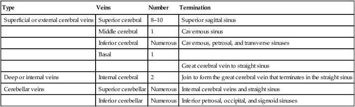
Type
Veins
Number
Termination
Superficial or external cerebral veins
Superior cerebral
8–10
Superior sagittal sinus
Middle cerebral
1
Cavernous sinus
Inferior cerebral
Numerous
Cavernous, petrosal, and transverse sinuses
Basal
1
Great cerebral vein to straight sinus
Deep or internal veins
Internal cerebral
2
Join to form the great cerebral vein that terminates in the straight sinus
Cerebellar veins
Superior cerebellar
Numerous
Internal cerebral veins and straight sinus
Inferior cerebellar
Numerous
Inferior petrosal, occipital, and sigmoid sinuses
![]()
Stay updated, free articles. Join our Telegram channel

Full access? Get Clinical Tree


Neurologic Vascular Procedures