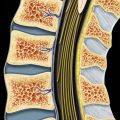
(Left) Sagittal graphic of a disc bulge shows generalized extension of the disc margin beyond the vertebral body . An axial view must show generalized extension > 90° of the disc margin to be a bulge; less than 90° would be a protrusion.
(Right) Sagittal graphic of a disc protrusion shows extension of disc material beyond the interspace margin where the base of the herniation is wider than the portion in the epidural space .
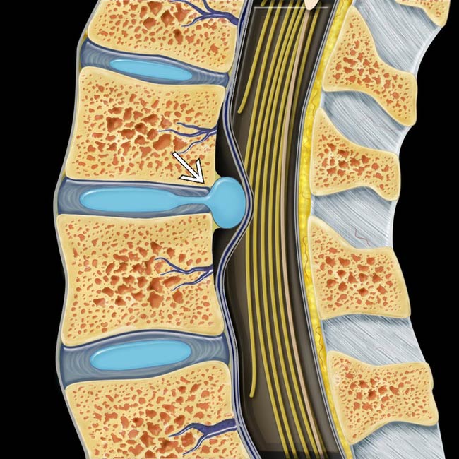
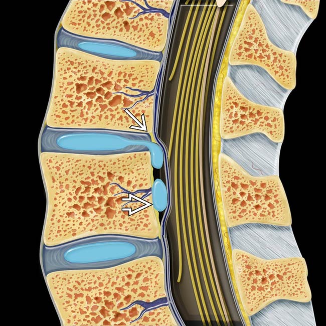
(Left) Sagittal graphic of a disc extrusion shows extension of disc material beyond the interspace where the base of the herniated material is smaller than the component in the epidural space .
(Right) Sagittal graphic shows a disc extrusion with free fragment. Extruded disc demonstrates extension of disc material beyond the interspace with the base narrower than the portion in the epidural space . A 2nd component has separated from the parent disc and is a free fragment or sequestered disc.
(Left) Sagittal T1WI MR shows severe disc degeneration at L5-S1 with disc extrusion where the base of herniation is narrower than the portion extending into epidural space . There is inferior migration of herniation consistent with free fragment.
(Right) Sagittal T1WI MR shows a large L3-L4 far lateral disc extrusion, which is located within the neural foramen (foraminal herniation) and obscures the exiting L3 nerve root. Note the normal exiting root at the level below for comparison .
Only gold members can continue reading. Log In or Register to continue
 . An axial view must show generalized extension > 90° of the disc margin to be a bulge; less than 90° would be a protrusion.
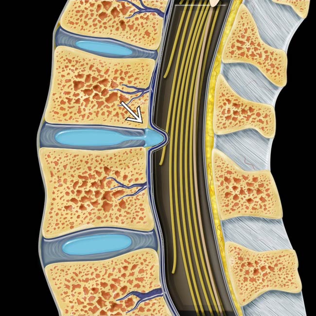
 .
.
 .
.
 . A 2nd component
  has separated from the parent disc and is a free fragment or sequestered disc.
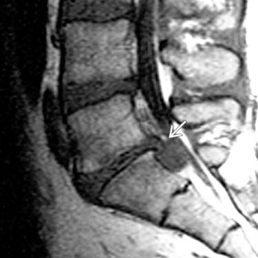
 . There is inferior migration of herniation consistent with free fragment.
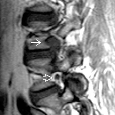
 and obscures the exiting L3 nerve root. Note the normal exiting root at the level below for comparison
  .
.