Usually unilocular, well-defined cyst with sharp margin and thin imperceptible wall
– Typically no internal complexity, septations, nodularity, or calcifications
– Usually no communication with pancreatic duct
– Usually single cyst but frequently multiple in patients with underlying syndrome
Less commonly, imaging features can overlap with neoplastic pancreatic cysts, and lesions can demonstrate more complexity (multiloculation, calcifications, etc.)
– Lymphoepithelial cysts are more commonly complex (may have macroscopic fat) and can be intrapancreatic, abut pancreas, or appear exophytic
May demonstrate signal loss on out-of-phase gradient-echo MR images (due to intracellular lipid)
May be connected with pancreas by tiny, imperceptible stalk and appear exophytic or extrapancreatic
• Syndromes account for most nonneoplastic cysts diagnosed prospectively in clinical practice
von Hippel-Lindau (VHL) disease, autosomal dominant polycystic kidney (ADPKD), and cystic fibrosis (CF)
• Isolated nonneoplastic cysts without a syndrome are far more rare in clinical practice
CLINICAL ISSUES
• ACR incidental findings committee suggests simple pancreatic cysts measuring ≤ 2 cm can be safely followed
• Simple pancreatic cysts in setting of a known syndrome (VHL, ADPKD, CF) are almost certainly benign
• Larger lesions or lesions with suspicious morphologic features often require EUS or cyst aspiration and consideration for surgical resection
(Left) Axial CECT in an asymptomatic patient demonstrates a simple-appearing, thin-walled cyst in the pancreatic neck.
(Right) Endoscopic ultrasound in the same patient shows a simple cyst in the neck of the pancreas. There is no mural nodularity or other sign of complexity. Aspiration demonstrated clear serous fluid with no elevated tumor markers. It was elected to follow this cyst with sonography. It has remained stable for several years and is presumably a nonneoplastic simple cyst.
(Left) Axial CECT shows a small, simple-appearing cyst in the pancreatic head/neck. The wall of the cyst is imperceptible and there are no internal septations or other signs of complexity.
(Right) Endoscopic ultrasound in the same patient confirms the simple appearance of the cyst . Needle aspiration of the cyst demonstrated clear serous fluid with no malignant cells or elevated tumor markers, consistent with a nonneoplastic, simple cyst.
TERMINOLOGY
Synonyms
• Congenital, true, or epithelial pancreatic cyst
Definitions
• Group of nonneoplastic, noninflammatory, benign pancreatic cysts comprising several different histopathologic entities
IMAGING
General Features
• Best diagnostic clue
Simple-appearing cyst with no septations or mural nodularity in a patient with no history of pancreatitis
– Consider strongly in patients with history of cystic fibrosis, autosomal dominant polycystic kidney disease (ADPKD), or von Hippel-Lindau (VHL)
• Size
Usually quite small, although rarely can be much larger: Giant cysts as large as 15 cm in diameter reported
• Morphology
Usually unilocular with round or oval shape, smooth thin wall, and absence of internal complexity
Solitary or multiple (when associated with cystic syndromes)
CT Findings
• Imaging features can show some variability, since this category encompasses several histopathologically distinct types of nonneoplastic cysts
Most nonneoplastic cysts are unilocular and well defined with a sharp margin and thin imperceptible wall
Typically no internal complexity, septations, nodularity, or calcifications
Usually no discernible communication with pancreatic duct
Usually single isolated cyst, but often multiple in patients with underlying syndrome
• Less commonly, imaging features can overlap with neoplastic pancreatic cysts, and lesions can demonstrate more complexity (multiloculation, calcifications, etc.)
Lymphoepithelial cysts have been described as more commonly demonstrating complexity (and even macroscopic fat) and may be either intrapancreatic, abut pancreas, or appear exophytic
– May be connected with pancreas by tiny, imperceptible stalk and appear exophytic or extrapancreatic
– Appear multilocular in 60% of cases
MR Findings
• Most nonneoplastic cysts are simple in appearance (hypointense on T1WI, hyperintense on T2WI, no enhancement or complexity)
Lesions may demonstrate more complexity and be indistinguishable from a cystic neoplasm
Lymphoepithelial cysts may demonstrate complexity and signal loss on out-of-phase gradient-echo images due to intracellular lipid
• Usually no communication with pancreatic duct on MRCP
Rarely, some histopathologic subtypes of nonneoplastic cysts (i.e., retention cysts) may communicate with pancreatic duct
Ultrasonographic Findings
• Most often anechoic with no internal complexity or echoes
Radiographic Findings
• ERCP: Usually no communication between cyst and duct
Imaging Recommendations
• Best imaging tool
CECT or MR followed by endoscopic US (EUS)
DIFFERENTIAL DIAGNOSIS
Pancreatic Pseudocyst
• More often complex in appearance with discrete wall
Only gold members can continue reading. Log In or Register to continue
 Less commonly, imaging features can overlap with neoplastic pancreatic cysts, and lesions can demonstrate more complexity (multiloculation, calcifications, etc.)


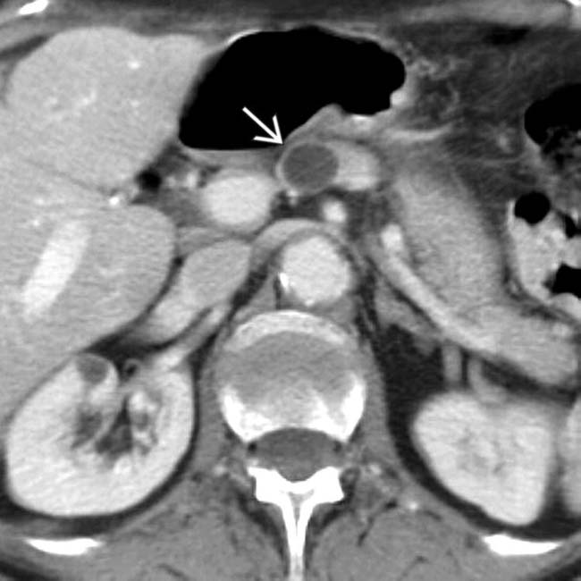
 in the pancreatic neck.
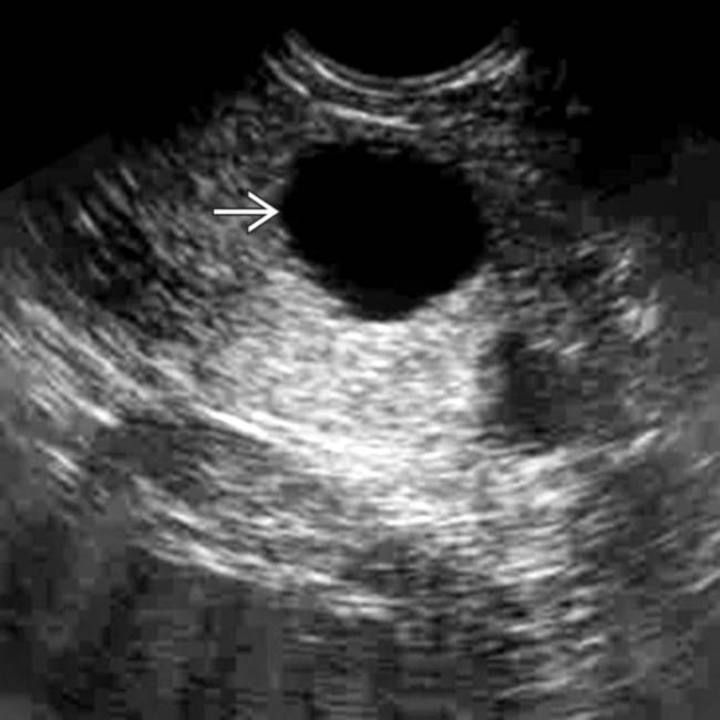
 in the neck of the pancreas. There is no mural nodularity or other sign of complexity. Aspiration demonstrated clear serous fluid with no elevated tumor markers. It was elected to follow this cyst with sonography. It has remained stable for several years and is presumably a nonneoplastic simple cyst.
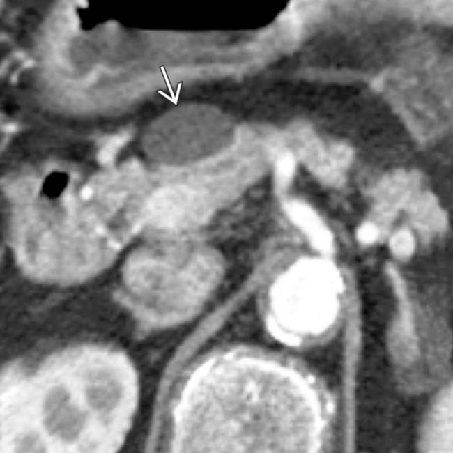
 in the pancreatic head/neck. The wall of the cyst is imperceptible and there are no internal septations or other signs of complexity.
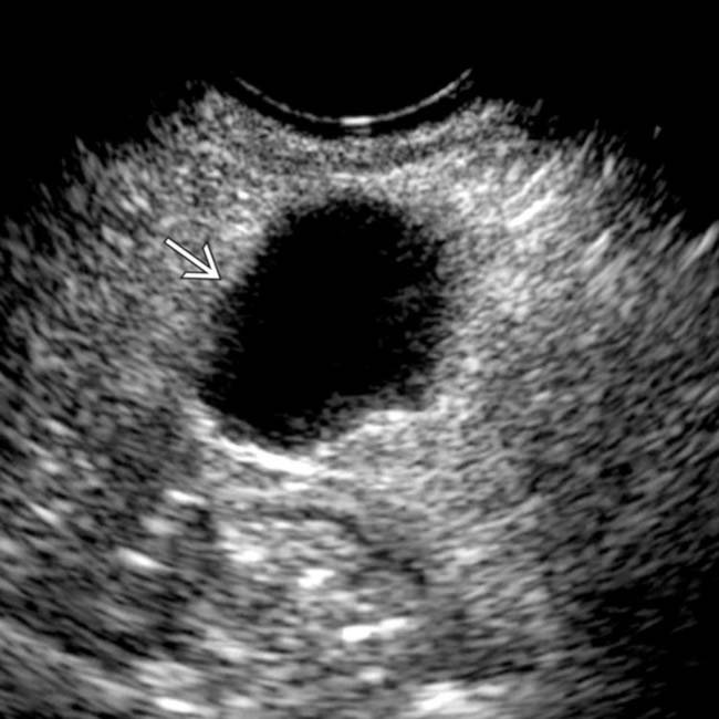
 . Needle aspiration of the cyst demonstrated clear serous fluid with no malignant cells or elevated tumor markers, consistent with a nonneoplastic, simple cyst.


 Most nonneoplastic cysts are unilocular and well defined with a sharp margin and thin imperceptible wall


