This article focuses on the variants and imaging pitfalls in the ankle and foot.
MR of the musculoskeletal structure
Great advances have been made in musculoskeletal radiology since the dawn of cross-sectional imaging more than three decades ago. Innovation in imaging technology has provided an unprecedented window into intra-articular pathology. MR imaging, in particular, has revolutionized the ability to study the anatomic details of all the components of the musculoskeletal system, including tendons, ligaments, muscles, and bones as well as the pathologic processes that affect them. Crucial to the accurate analysis of these structures is a solid knowledge of the anatomic variants that can be misinterpreted for pathology on MR imaging. This article focuses on the variants and imaging pitfalls in the ankle and foot.
Technical factors
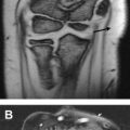
As a general rule, the tendons and ligaments of the ankle and foot, due to their highly organized architecture and collagen composition, demonstrate homogeneously hypointense signal on all pulse sequences. Loss of the expected low intrasubstance signal in a tendon or ligament is considered the hallmark in the diagnosis of pathologic conditions. The magic angle phenomenon is a technical phenomenon that can mimic pathology. The magic angle effect occurs when the orientation of the collagen fibers approximates the magic angle of 55° with the main magnetic vector (Z axis). This phenomenon is particularly prominent when a low echo time (TE) of 10 to 20 milliseconds is used, as in T1-weighted, proton density, and gradient-echo sequences. T2-weighted or short tau inversion recovery (STIR) images with high TE values (>35 milliseconds) eliminate the magic angle effect. When imaging the ankle, this phenomenon can be reduced by scanning patients in the prone position or positioning the foot at 20° of plantar flexion while scanning patients in a supine position. The magic angle more frequently affects the posterior tibial tendon just proximal to its navicular insertion, the peroneal tendons in subfibular position, and the anterior tendons at the level of the ankle joint ( Figs. 1 and 2 ).


Incomplete fat suppression can falsely produce hyperintense signal in the soft tissues and bone marrow, most commonly in the lateral aspect of the ankle in the region of the lateral malleolus, although the medial malleolus can also be affected ( Fig. 3 ). This is thought to be due to factors, such as coil proximity artifact and the presence of inhomogeneities in the static magnetic field. Inhomogeneous fat saturation can be combated through the use of inversion recovery imaging, which is insensitive to field inhomogenities, and by the use of multichannel phase array coils.

Technical factors
As a general rule, the tendons and ligaments of the ankle and foot, due to their highly organized architecture and collagen composition, demonstrate homogeneously hypointense signal on all pulse sequences. Loss of the expected low intrasubstance signal in a tendon or ligament is considered the hallmark in the diagnosis of pathologic conditions. The magic angle phenomenon is a technical phenomenon that can mimic pathology. The magic angle effect occurs when the orientation of the collagen fibers approximates the magic angle of 55° with the main magnetic vector (Z axis). This phenomenon is particularly prominent when a low echo time (TE) of 10 to 20 milliseconds is used, as in T1-weighted, proton density, and gradient-echo sequences. T2-weighted or short tau inversion recovery (STIR) images with high TE values (>35 milliseconds) eliminate the magic angle effect. When imaging the ankle, this phenomenon can be reduced by scanning patients in the prone position or positioning the foot at 20° of plantar flexion while scanning patients in a supine position. The magic angle more frequently affects the posterior tibial tendon just proximal to its navicular insertion, the peroneal tendons in subfibular position, and the anterior tendons at the level of the ankle joint ( Figs. 1 and 2 ).
Incomplete fat suppression can falsely produce hyperintense signal in the soft tissues and bone marrow, most commonly in the lateral aspect of the ankle in the region of the lateral malleolus, although the medial malleolus can also be affected ( Fig. 3 ). This is thought to be due to factors, such as coil proximity artifact and the presence of inhomogeneities in the static magnetic field. Inhomogeneous fat saturation can be combated through the use of inversion recovery imaging, which is insensitive to field inhomogenities, and by the use of multichannel phase array coils.
Tendons
Anterior Compartment
The anterior compartment tendons (anterior tibial, extensor hallucis longus, extensor digitorum longus, and peroneus tertius) are rarely injured. The anterior tibial tendon is the most commonly injured extensor tendon. Tear of the anterior tibial tendon is classically seen in older patients and athletes who run hills. Apparent longitudinal split tearing in the insertional portion of this tendon at the medial cuneiform bone and base of the first metatarsal in asymptomatic patients is most likely related to the presence of multiple insertional slips. This is thought to represent a normal variant.
Medial Compartment
The insertional portion of the posterior tibial tendon on the medial navicular tubercle typically has a heterogenous appearance. This signal heterogeneity is secondary to a combination of magic angle effect and fat interposed between the insertional slips of the tendon. Another cause of the heterogeneity is the presence of an intratendinous accessory navicular bone (type I accessory navicular bone [os tibiale externum]) ( Fig. 4 ).
A small amount of tenosynovial fluid is frequently observed within the tendon sheaths in asymptomatic individuals and should not be considered abnormal. Physiologic tenosynovial fluid is more frequently found in the flexor than in the extensor tendons. The lack of tendon sheath in the distal preinsertional portion of the posterior tibial tendon renders fluid signal in this location abnormal, however. Posterior tibial peritendinitis likely related to metaplastic synovium should be considered. Disproportionate fluid within the flexor tendon sheaths, compared with the amount of fluid found in the ankle joint, is usually indicative of tenosynovitis. Communication between the ankle joint and the flexor hallucis longus tendon, however, explains the presence of prominent fluid within this tendon sheath in patients with large joint effusions. Thus, even a large amount of fluid within the tendon sheath of the flexor hallucis longus tendon may be of no clinical significance.
Posterior Compartment
The tendons of the gastrocnemius and soleus muscles form the Achilles tendon. The Achilles tendon measures approximately 15 cm in length and typically has a fascicular appearance secondary to the intermixing of its fibers with fibrofatty tissue and vessels. This, in turn, produces intrasubstance linear and punctate hyperintense foci on T1-weighted and gradient-echo images. Preservation of the normal morphology of the tendon helps differentiate this signal heterogeneity from a partial tear because a normal tendon maintains its normal flat/concave shape anteriorly and demonstrates intact fibers throughout its course without intervening fluid-like T2 signal. In addition, the fascicular appearance is usually less apparent on STIR and T2-weighted images.
Low or incomplete incorporation of the gastrocnemius and soleus tendons may produce heterogeneity due to persistent fat planes between the tendon slips. Assessment of the course of the soleus tendon relative to the gastrocnemius tendon on sequential axial images avoids confusing this normal variant with disease ( Fig. 5 ). A pathologic process that can mimic this appearance is a xanthomatous Achilles tendon. Xanthomas are composed of lipid-filled foamy histiocytes and extracellular cholesterol deposits. They are typically seen in patients with inherited metabolic diseases, such as familial hypercholesterolemia and hyperproteinemia. A xanthomatous Achilles tendon typically has a speckled or reticulated appearance with or without tendon enlargement. This MR imaging appearance correlated with a patient’s medical history helps diagnose this pathologic process.
Lateral Compartment
The peroneal tendons (peroneus brevis and peroneus longus) share a common tendon sheath down to the level of the lateral malleolar tip. From this point on, the tendons have individual tendon sheaths. The peroneal tendons can be affected by magic angle effect because they course obliquely around the lateral malleolus and onto their insertions in the foot, particularly as the peroneus longus tendon curves beneath the cuboid bone. The primary restraints to subluxation of the peroneal tendons are the superior and inferior peroneal retinacula. The superior peroneal retinaculum courses from the posterolateral aspect of the distal fibula to the lateral calcaneus and helps stabilize the tendons in the retromalleolar groove. The inferior peroneal retinaculum attaches to the peroneal trochlea and calcaneus above and below the tendons while forming a septum that reinforces the individual tendon sheaths. The oblique course of the peroneus brevis tendon leads to apparent subluxation of the tendon where the brevis tendon is found medial to the medial margin of the fibular groove instead of at its more frequent position posterior to the fibular groove and anterior to the peroneus longus tendon. Pseudosubluxation of the peroneus brevis tendon can be further accentuated in foot supination.
Ligaments
Lateral Compartment
Of the three main ligaments in the low lateral compartment of the ankle, the least prone to injury is the posterior talofibular ligament. This ligament tends to have a fan-like, striated appearance on MR imaging that should not be confused with a sprain or tear. The posterior talofibular ligament and the posterior intermalleolar ligament course transversely posterior to the tibiotalar joint and, thus, frequently appear as punctuate hypointensities as they are imaged in cross section in the sagittal plane. This appearance can mimic posterior ankle intra-articular bodies. It is important to carefully track each of these ligaments from their origin to insertion to exclude the presence of a loose body using the orthogonal imaging planes.
Syndesmotic Ligaments
The anterior tibiofibular ligament can also pose a diagnostic dilemma, because it may appear thickened and discontinuous. This appearance can be a normal finding and is thought to be related to fat interposed between the fibers that make up the ligament as well as due to its downward oblique orientation from the anterior tibial lip to its insertion into the fibular malleolus ( Fig. 6 ). As with the posterior intermalleolar and posterior talofibular ligaments (discussed previously), the anterior and posterior tibiofibular ligaments can mimic intra-articular loose bodies on the sagittal plane due to their transverse course.
Medial Compartment
The deltoid ligament complex is made up of superficial and deep layers. One of the most readily visualized components of the complex is the posterior tibiotalar ligament, a component of the deep layer. This ligament has a striated appearance due to the presence of intervening fat between its fibers, which should not be confused with injury. This striated appearance is regularly seen in the young adult population.
The spring ligament complex is made up of three separate components: the superomedial, the medial plantar oblique, and the inferior plantar longitudinal fibers. The spring ligament complex is an important structure of the anterior subtalar joint providing a fibrocartilaginous articular surface to the talar head ( Fig. 7 A ). The superomedial component, which courses from the medial aspect of the sustentaculum tali to the superomedial aspect of the navicular, runs medial to the distal posterior tibial tendon and is separated by loose connective tissue. This loose connective tissue helps differentiate between the posterior tibial tendon and superomedial spring ligament. Frequently noted between the medial plantar oblique and the inferior plantar longitudinal ligaments is a fluid-filled recess of the talocalcaneonavicular ( Fig. 7 B). Fluid signal within this recess may potentially be misconstrued as a spring ligament tear. Visualization of the recess is facilitated by the presence of a native effusion or by intra-articular injection of contrast solution in the talonavicular joint. Posttraumatic talonavicular effusions in the setting of acute impaction injury of the talar head and talonavicular osteoarthritis are often seen in association with a fluid distended spring recess.
Accessory Ligaments
The posterior intermalleolar ligament is a ligament found in the posterior aspect of the ankle in 81.8% of specimens. It was originally described as a ligamentous structure extending between the medial malleolus and the lateral malleolus ( Fig. 8 ). Recent studies have reported a diverse group of medial origins with the lateral insertion consistently found in the medial fossa of the lateral malleolus. This ligament can have different shapes depending on the site of its medial origin as well as of the number of fiber bundles and their density. The posterior intermalleolar ligament can potentially become entrapped and be a cause of posterior ankle impingement.
In general, it is important to correlate the clinical history along with the imaging findings when evaluating these structures for injury. The morphology of each ligament and tendon should be carefully examined as well as its signal in conjunction with the status of the surrounding soft tissue and osseous structures to confidently differentiate a tear or sprain from a normal variant.
Muscles
Muscle variants are frequently seen in the ankle. These muscles are usually asymptomatic and often incidentally found on MR imaging obtained for unrelated reasons. When they do come to attention, they can either present as a mass on physical examination or cause pain related to their effect on the surrounding structures. For instance, the peroneal tunnel can house an accessory muscle, the peroneus quartus, which is located adjacent to the peroneus brevis and peroneus longus tendons. This muscle can cause peroneal tunnel overcrowding, leading to mass effect and compression on the peroneus brevis and peroneus longus tendons and eventual tearing. The peroneus quartus inserts independently into the retrotrochlear eminence of the lateral calcaneus, which helps differentiate it from a low-lying peroneus brevis muscle belly ( Fig. 9 ). The latter, unlike the peroneus quartus, does not predispose to tearing. The flexor accessorius digitorum longus (FADL) is the most common accessory muscle found within the tarsal tunnel. Due to its location inside the tarsal tunnel, the FADL can cause mass effect on the adjacent tibial nerve or its plantar branches, particularly during exercise-related engorgement leading to neuropathy and denervation ( Fig. 10 ). Other common normal muscle variants include the accessory soleus, low incorporation of the soleus, and peroneo calcaneus internus muscles. An accessory soleus can be distinguished from low incorporation of the soleus by following the course of the tendon in question. The tendon of the accessory soleus inserts separately onto the calcaneus anteromedial to the attachment of the Achilles.
Related posts:
 Elbow Magnetic Resonance Imaging Variants and Pitfalls
 Pitfalls of Wrist MR Imaging
 MR Imaging of the Hip: Normal Anatomic Variants and Imaging Pitfalls
Magnetic Resonance Imaging of the Midfoot and Forefoot: Normal Variants and Pitfalls
Bone Marrow
MR Imaging of the Hip: Normal Anatomic Variants and Imaging Pitfalls
Magnetic Resonance Imaging of the Midfoot and Forefoot: Normal Variants and Pitfalls
Bone Marrow
 MR Imaging Features of Common Variant Spinal Anatomy
Stay updated, free articles. Join our Telegram channel

Full access? Get Clinical Tree


