Case presentation
A 9-year-old male presents with a 4-month history of right upper arm pain. Over the past 2 months, he and his parents have noted the development of swelling to the arm as well. The patient is active in sports, particularly baseball, and the pain started after he pitched a game. He was given ibuprofen with some relief of the pain, but over the past several weeks, this has not been helping. He has not had fever, numbness, weakness, rash, or other symptoms.
Physical examination reveals a well-appearing pleasant patient in no acute distress. His vital signs are unremarkable. He has a normal physical examination with the exception of his right upper arm, where there is swelling, and tenderness of the midshaft of the right humerus. While he has normal gross range of motion, he does complain of pain with movement of the arm. There is no erythema, warmth, or fluctuance. He is neurologically intact.
Imaging considerations
Plain radiography
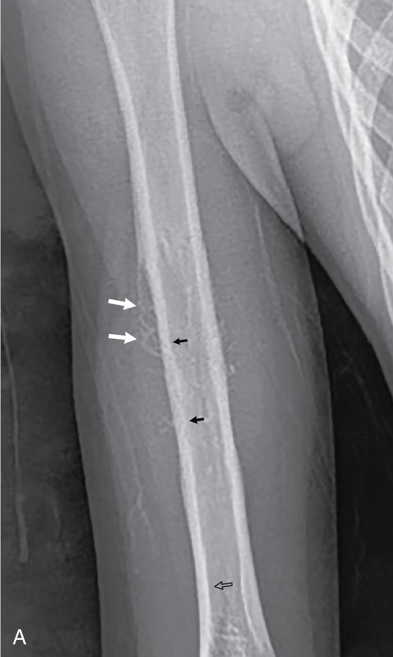
Plain radiography is the initial imaging modality of choice in patients with musculoskeletal complaints. Since Ewing sarcoma commonly presents with pain, this modality is the first-line modality utilized in these patients. A two-view imaging study (anteroposterior and lateral) is usually obtained. This study may reveal osteolysis and periosteal reaction. The periosteal reaction of Ewing sarcoma usually reflects the aggressive nature of the lesion and can produce characteristic imaging findings such as the classically described “onion skin” periosteal new bone and the presence of the “Codman triangle.” With an aggressive lesion, the periosteal reaction is often discontinuous, as the tumor growth is often too rapid to allow for smooth, continuous periosteal reaction. Instead, the periosteal reaction will be seen to abruptly terminate, forming a Codman triangle, as seen subsequently in this patient and in Chapter 78 ( Fig. 78.1 ). The Codman triangle is an area of new subperiosteal bone created when the tumor raises the periosteum away from the bone and is often accompanied by spicule formation in the soft tissues. ,
Computed tomography (CT)
This imaging modality may be appropriate as a secondary study when initial plain radiography is abnormal; however, magnetic resonance imaging (MRI) is the most appropriate secondary imaging study for a suspected malignant primary bone tumor. CT may be used in the staging process, including evaluation of metastasis to the lungs, once the diagnosis of Ewing sarcoma is suspected. , The rapidity and wide availability of this imaging test, as well as the lack of sedation need, are advantages for CT.
Magnetic resonance imaging
MRI with and without intravenous contrast is the advanced imaging modality of choice when initial plain radiography is abnormal or unremarkable, and a primary bone tumor is otherwise suspected. MRI is excellent at defining intramedullary involvement and depicting the soft tissue extent of a lesion. , Since this modality is also very good at delineating the anatomic relationships between the tumor and surrounding structures, it is useful at planning operative intervention and biopsies. , ,
Positron emission tomography (PET)
This modality is used as a follow-up imaging test in the staging and treatment process; it is not a first-line imaging modality.
Imaging findings
The patient’s history and physical examination findings warrant further investigation. Plain radiography of the right humerus was obtained. These images were somewhat difficult to obtain, since the patient had pain with movement of the arm despite pain medication. These images revealed a destructive lesion in the midshaft of the right humerus, with a permeative lytic pattern in the cortex. The periosteal reaction surrounding the lesion was indicative of an aggressive lesion, with a Codman triangle and spiculated periosteal new bone (see Chapter 78 [Osteosarcoma], for further discussion of the Codman triangle). These patterns occur as an aggressive tumor lesion that expands beyond the bone, elevates the periosteum, and lesion growth outpaces the growth of newly-formed subperiosteal bone ( Fig. 79.1 ).
Related posts:


Stay updated, free articles. Join our Telegram channel

Full access? Get Clinical Tree


