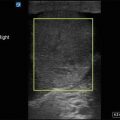
Figure 12.1
Transabdominal transverse gravid uterus. Using a transabdominal approach, the gravid uterus in visualized here in a transverse plane. The fetus, in this case, is lying in a sagittal plane
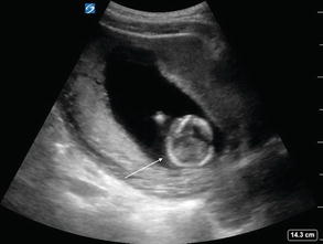
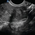
Figure 12.2
Transabdominal uterus in sagittal. Using a transabdominal approach, the gravid uterus is visualized here in a sagittal view. The fetal head is a hyperechoic structure within hypoechoic fluid (arrow)
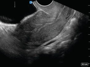
Figure 12.3
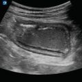
Bladder with uterus in sagittal plane. In this non-gravid uterus, the uterine stripe can be seen in the midline of the uterus (horizontal arrows) with the bladder noted to the left (vertical arrow). This uterus is anteverted, the most common uterine position

Figure 12.4
Transvaginal gravid uterus, sagittal. Using a transvaginal approach, the gravid uterus is visualized in a sagittal plane. Note the hypoechoic circular structure in the midline of the uterine stripe within the uterine fundus. This represents a gestational sac containing a yolk sac
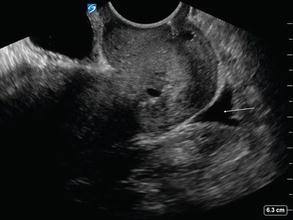
Figure 12.5
Pouch of Douglas. The pouch of Douglas, also referred to as the cul-de-sac or rectouterine pouch, lies posterior to the uterus and anterior to the rectum (arrow) seen in this sagittal transvaginal view. Trace to small amount of physiologic free fluid is not unusual in women of childbearing age
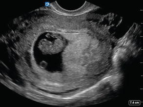
Figure 12.6
Transvaginal gravid uterus, transverse. Using a transvaginal approach, the gravid uterus is visualized in a transverse plane with a large gestational sac on the left of the image. A yolk sac and developing fetus are clearly visible
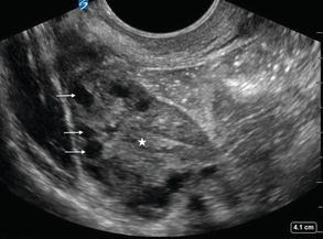
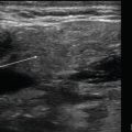
Figure 12.7
Normal ovary. Multiple small cysts (arrows) in the cortex surround the medulla (star) in this normal ovary, giving the appearance of what is often referred to as a “chocolate chip cookie”
Evaluation for Intrauterine Pregnancy
- (a)
Gestational Sac
A gestational sac can be identified at approximately 5 weeks of gestation [3] and in the majority of patients with a bHCG greater than 1000–2000 mIU/mL [1–3].
It will appear as a round or oval hypoechoic fluid collection within the uterine stripe, typically in the fundus:
Figure 12.8—Empty gestational sac
Video 12.5—Empty gestational sac
This can be present in a normal pregnancy prior to development of a yolk sac or fetal pole within the intrauterine gestation sac.
However, this also can be seen with an ectopic pregnancy, termed pseudogestational sac.
The presence of a gestational sac without a yolk sac and fetal pole does not confirm intrauterine pregnancy, especially in cases where ectopic pregnancy is suspected.
- (b)
Double Decidual Sac
Visualized as alternating hyperechoic rings separated by a thin hypoechoic layer of fluid surrounding a gestational sac:
The inner ring is called the decidua capsularis.
The outer ring is called the decidua parietalis.
The presence of a double decidual sac without a yolk sac and fetal pole is not confirmatory of an intrauterine pregnancy.
Figure 12.9—Double decidual sac.
Video 12.6—Double decidual sac.
- (c)
Yolk Sac
Located within a gestational sac.
Will appear as a small, thin-walled, spherical structure with an anechoic center:
Figure 12.10—Yolk sac
Video 12.7—Yolk sac
This is the first confirmatory sign of true intrauterine pregnancy [3].
Usually appears at approximately 5–6 weeks of gestation and disappears by 12 weeks [3].
- (d)
Fetal Pole
- (e)
Fetal Heart Rate
- (f)
Gestational Age Measurements
There are several different measurements that can be done to estimate gestation age. However, the easiest to perform are crown-rump length (CRL) and biparietal diameter (BPD).
Gestational age measurements are most accurate in first trimester.
Crown-rump length:
This measurement can be obtained at about 6 weeks of gestation [3].
Image the fetus to visualize the spine in a longitudinal plane.
Use calipers to measure from the top of fetal head to bottom of the buttocks:
Figure 12.13—Crown-rump length
Use this measurement to compare with standardized CRL-to-gestational age charts or, if available, use built in software to determine gestational age.
Biparietal diameter:
This measurement can be obtained toward the end of the first trimester and within the second trimester [3].
Obtain a transverse view of the fetal head with the thalami visible and symmetric.
Use calipers to measure from the outer edge of one bony table to the inner edge of the other [3], across the widest portion.
Figure 12.14—Biparietal diameter
Use this measurement to compare with standardized BPD-to-gestational age charts or, if available, use built in software to determine gestational age.
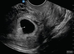
Figure 12.8
Empty gestational sac. An empty gestational sac (arrow) is visualized here within the uterus. This is the first early sign of a developing pregnancy, but does not in itself confirm an intrauterine pregnancy
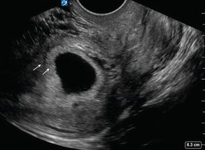
Figure12.9
Double decidual sign. Note the alternating hyperechoic rings separated by a thin hypoechoic layer of fluid surrounding a gestational sac
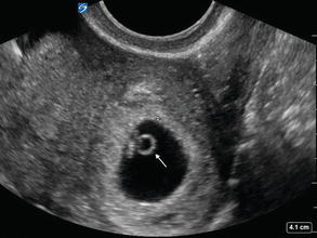
Figure 12.10
Yolk sac. A yolk sac (arrow) is the first true ultrasonographic confirmation of intrauterine pregnancy
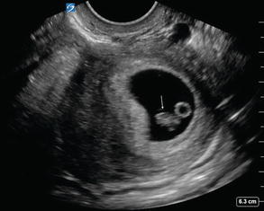
Figure 12.11


Fetal pole. A fetal pole (arrow) first appears as a thickened area on the yolk sac at approximately 6 weeks of gestational age by transvaginal ultrasound. The circular yolk sac is also visualized
Related posts:
Stay updated, free articles. Join our Telegram channel
Full access? Get Clinical Tree
