42 Obturator Nerve Block
The obturator nerve arises from the lumbar plexus and innervates most of the adductors of the medial compartment of the thigh. The other adductors are the pectineus (innervated by the femoral nerve) and the adductor magnus (partially innervated by the sciatic nerve). The abundance of motor fibers makes the obturator nerve a frequent choice for electromyographic recording of compound motor action potentials (CMAPs).1
The cutaneous innervation by the obturator nerve is variable.2 However, there are morphine-sparing effects of obturator nerve block after major surgical procedures of the lower extremity. In blocks in which multiple lower extremity nerves are targeted (e.g., the posterior lumbar plexus block or the anterior 3-in-1 block), the obturator nerve has a relatively low block success rate. Therefore, obturator nerve block is an important adjunct for lower extremity analgesia. Other indications for obturator nerve block include relief of hip pain, treatment of adductor spasticity, and prevention of obturator stimulation during transurethral resection of lateral bladder wall tumors. Change in adduction strength is the best method for assessing obturator nerve block. However, even with complete obturator nerve block, there is some residual adduction strength because the pectineus (femoral nerve innervation) and the hamstring component of the adductor magnus (sciatic nerve innervation) muscles remain intact.
Suggested Technique
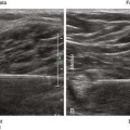
The anterior and posterior divisions of the obturator nerve converge proximally along the rounded lateral border of the adductor brevis muscle. The obturator nerve divisions are thin and flat as the fascicles disperse to the muscle groups. The obturator nerve divisions have a “white bands” appearance on sonography.3 It is important that the flat surfaces of the obturator nerve divisions are perpendicular to the sound beam to enhance their echo brightness. Note that although the anterior and posterior divisions converge along the lateral border of the adductor brevis, they do not actually meet there in most (75%-80%) subjects because the divisions remain separated by the obturator externus muscle proximally.4 Therefore, the obturator nerve block is usually performed as a multiple-injection technique targeting each of the two divisions separately.


Stay updated, free articles. Join our Telegram channel

Full access? Get Clinical Tree


