Histo: fine connective tissue + cellular septa link pia and arachnoid
› contains CSF that drains through the valves of arachnoid granulations into venous sinuses
› forms basal cisterns
3. Pia mater [ pia , Latin = tender]
= delicate innermost layer of meninges
Histo: thin fibrous tissue impermeable to fluid with perforations for blood vessels to pass through
F. SUBPIAL SPACE
= perivascular space = VR (Virchow-Robin) space
[Rudolf Virchow (1821–1902), pathologist in Berlin, Germany]
[Charles P. Robin (1821–1885), anatomist in Paris, France]
Histo: no communication with subarachnoid space; VR space around intracortical artery continues within subarachnoid space; VR space around cerebral vein is continuous with subpial space
Function: lymphatic drainage system of the brain
| Sites: | Type I | = | lenticulostriate arteries |
| Type II | = | medullary arteries over high convexities | |
| Type III | = | collicular arteries in midbrain |
√ smoothly demarcated typically < 5 mm fluid-filled cyst; often in clusters
√ SI visually similar to CSF (actually lower when measured as VR spaces are entrapments of interstitial fluid)
√ no restricted diffusion + no enhancement
√ inflow effects on flow-sensitive T1WI
G. EPENDYMA
= thin epithelial-like lining of ventricular system + central canal of spinal cord composed of ciliated simple columnar ependymocytes
Origin: one of four types of neuroglia in CNS
Function: (1) Production + regulation of CSF, (2) Reservoir for neuroregeneration
Falx Cerebri [falx, Latin = curved blade or scythe]
= large crescent-shaped inelastic reflection of meningeal layer of dura mater that descends vertically in longitudinal fissure between cerebral hemispheres
Connected to:
(a) anteriorly: crista galli anteriorly in proximity to cribriform plate + frontal and ethmoid sinuses
(b) posteriorly: upper surface of the tentorium cerebelli
Margins:
(a) superior margin attached at midline to internal surface of skull as far back as internal occipital protuberance
› contains superior sagittal sinus overlying longitudinal cerebral fissure
(b) inferiorly adjacent to corpus callosum + cingulate gyrus + pericallosal arteries
› contains inferior sagittal sinus arching over corpus callosum deep in longitudinal cerebral fissure
Falx Cerebelli
= small sickle-shaped fold of dura mater projecting forward into posterior cerebellar notch + into cerebellar vallecula between cerebellar hemispheres
Base: attached to inferoposterior part of tentorium cerebelli
Posterior margin: attached to vertical crest of inner skull surface below internal occipital protuberance
CLASSIFICATION OF BRAIN ANATOMY
A. PROSENCEPHALON = forebrain
forms from process of ventral induction (ie, 3 closely interconnected sequential events of formation + cleavage + midline development)
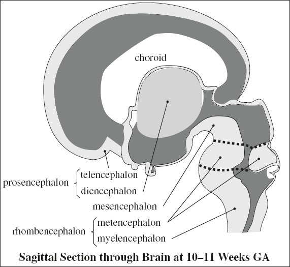
√ cerebrum, lateral ventricles, choroid, thalami, cerebellum sonographically visible at 12 weeks MA
1. Telencephalon = cerebrum
= cerebral hemispheres, putamen, caudate nucleus
2. Diencephalon
= thalamus, hypothalamus, epithalamus (= pineal gland + habenula), globus pallidus, optic vesicles
B. MESENCEPHALON = midbrain
= short segment of brainstem above pons; traverses the hiatus in tentorium cerebelli; contains cerebral peduncles, tectum, colliculi (corpora quadrigemina)
C. RHOMBENCEPHALON = hindbrain
√ posterior cystic space of 4th ventricle sonographically detectable between 8 and 10 weeks MA
1. Metencephalon = cerebellar hemispheres, vermis
2. Myelencephalon = medulla oblongata, pons
D. BRAINSTEM = midbrain + pons + medulla contains
(a) cranial nerve nuclei
(b) sensory and motor tracts between thalamus, cerebral cortex, and spinal cord
(c) reticular formation controlling respiration, blood pressure, gastrointestinal function, centers for arousal and wakefulness
THALAMUS
= midline structure situated between cerebral hemispheres + midbrain with paired symmetric portions
Location: on either side of 3rd ventricle
Function: relay of sensory + motor signals; regulation of consciousness, sleep, alertness
Blood supply: PCA + pCom
Hypothalamus [hypo, Greek = below; thalamus, Greek = bed]
= part of diencephalon below the thalamus
Origin: neuroectoderm
Boundaries:
(a) anterior = lamina terminalis extending from anterior commissure to optic chiasm
(b) posterior = line extending from mamillary bodies to posterior commissure
(c) lateral = medial thalamus
(d) inferior = tuber cinereum (posteriorly) + median eminence (middle) + infundibular stalk (anteriorly)
Function: homeostasis for blood pressure, body temperature, fluid + electrolyte balance, body weight
Regulatory mechanism:
(a) endocrine secretion:
› neuronal stimulation of posterior pituitary gland via infundibulum
› hypothalamic releasing factors to anterior pituitary gland via portal plexus as a vascular conduit
(b) autonomic function
(c) emotions
BASAL NUCLEI
= BASAL GANGLIA (earlier incorrect designation)
Function: part of extrapyramidal motor system; involvement in memory, emotion, other cognitive function
Blood supply: ACA and MCA → medial + lateral lenticulostriate aa.
A. Amygdaloid body
B. Claustrum
C. Corpus striatum
Location: between lateral ventricle + insular cortex
| Cranial Nuclei of Brainstem and Reticular Formation | ||
| A | = | sleep, wakefulness, consciousness |
| B | = | visual spatial orientation, higher autonomic coordination of food intake |
| C | = | pneumotaxic center, coordination of breathing and circulation |
| D | = | swallowing |
| E | = | blood pressure, cardiac activity, vascular tone |
| F | = | expiration |
| G | = | area postrema = trigger zone for vomiting |
| H | = | inspiration |
(1) Caudate nucleus
√ isointense to cortical gray matter on all pulse sequences
√ no enhancement
(2) Lentiform nucleus
√ may exhibit dilated Virchow-Robin perivascular spaces
√ may contain bilateral symmetric age-related calcifications
(a) pallidum = globus pallidus
√ slightly hypointense relative to putamen ← progressive iron deposition with age
(b) putamen
√ isointense to cortical gray matter on all pulse sequences
√ no enhancement
PITUITARY GLAND
= hypophysis cerebri within hypophyseal fossa of sphenoid, covered superiorly by sellar diaphragm (= dura mater) which has an aperture for the infundibulum centrally
Size: (adult size is achieved at puberty)
Height in adult females: 7 (range 4–10) mm
Height in adult males: 5 (range 3–7) mm
(normal height in men aged 40–49 = 4.89 ± 0.87 mm)
Shape:
√ flat / downwardly convex superior border
√ upwardly convex during puberty, pregnancy, in hypothyroidism (due to hyperplasia)
Anterior Lobe of Pituitary Gland
= larger anterior portion of adenohypophysis comprising 80% of pituitary gland volume
Origin: ectodermal derivative of stomadeum
Function:
(a) chromophil cells
1. acidophil cells = α cells
› growth hormone = somatotropin (STH)
› prolactin = lactogenic hormone (LTH)
2. basophil cells = β cells
› adrenocorticotropic hormone (ACTH)
› thyrotropin = thyroid-stimulating hormone (TSH)
› follicle-stimulating hormone (FSH)
› interstitial-cell-stimulating hormone (ICSH)
› luteinizing hormone (LH)
› melanocyte-stimulating hormone (MSH)
(b) chromophobe cells
50% of epithelial cell population, of unknown significance
MR:
√ larger homogeneous component isointense to white matter on T1WI + T2WI
√ prominent contrast enhancement (during first 3 minutes) ← lack of blood-brain barrier
√ hyperintense in newborn fading to normal adult signal by 2nd month of life
Pars Intermedia of Pituitary Gland
= posterior portion of adenohypophysis, separated from anterior lobe by hypophyseal cleft in fetal life
Origin: Rathke cleft / Rathke pouch within intermediate lobe of pituitary gland
Function: termination point of short hypothalamic axons elaborating tropic hormones (= releasing factors + prolactin inhibiting factor), which are carried to anterior lobe via the portal system
√ not visible with imaging techniques
Posterior Lobe of Pituitary Gland
= major portion of neurohypophysis


= termination point of neurosecretory axons from supraoptic and paraventricular nuclei of hypothalamus (= hypothalamohypophyseal tract)
Origin: diencephalic outgrowth
Function: storage site for transported
› vasopressin (= antidiuretic hormone [ADH])
› oxytocin
MR:
√ crescent of T1-hyperintensity + T2-isointensity compared with anterior pituitary lobe ← lipid in glial cell pituicytes + phospholipids of vasopressin
√ isointense in 10% of normal individuals
Pituitary Stalk / Infundibulum
Origin: arises from anterior aspect of floor of 3rd ventricle (infundibular recess)
Histo: formed from axons of cells lying in supraoptic + paraventricular nuclei of hypothalamus
√ joins posterior lobe at junction of anterior + posterior lobes
√ up to 3 mm thick superiorly, up to 2 mm thick inferiorly
√ usually in midline, may be slightly tilted to one side
MR:
√ prominent contrast enhancement
SEPTUM PELLUCIDUM
= 1.5–3.0 mm thin vertical membrane of triangular shape (when viewed from side)
› connecting corpus callosum to columns of fornix
› forming the medial wall of the lateral ventricles
› separating both anterior horns
Base of triangle: located anteriorly
Apex of triangle: located posteriorly
Five layers (leaves / laminae) of septum:
› CSF in anterior horn of RT lateral ventricle
› ependymal lining (gray matter) of RT lamina
› pial layer (white matter) of RT lamina
» potential space / slitlike cavity / cavum
› pial layer (white matter) of LT lamina
› ependymal lining (gray matter) of LT lamina
› CSF in anterior horn of LT lateral ventricle
Borders:
below: column + body of fornix
above: corpus callosum
ventral: continuous with precommissural septum + subcallosal gyrus
lateral: wall of lateral ventricles
Function: ?, part of limbic system moderating rage + arousal
Cavum Septi Pellucidi
= persistence / failure of fusion of potential space (= slitlike cavity = 5th ventricle) between the 2 laminae forming septum pellucidum
Frequency: in 80% of term infants; in 15% of adults
Location: posterior to genu of corpus callosum, inferior to body of corpus callosum, anterosuperior to anterior pillar of fornix
Marker for: cerebral dysfunction
√ extends to foramen of Monro
√ may dilate + cause obstructive hydrocephalus (rare)
Regression: from back to front as fetus approaches term / in first few weeks after birth
Cavum Vergae
= “6th ventricle”
[Andrea Verga (1811–1895), clinical professor of psychiatry at Ospedale Maggiore in Milan, Italy]
= fluid-filled cavity located posterior to a vertical plane formed by columns of fornix
Frequency: in 30% of term infants; in 15% of adults
Location: posterior to fornix; anterior to splenium of corpus callosum inferior to body of corpus callosum superior to transverse fornix
√ posterior midline continuation of cavum septi pellucidi beyond foramen of Monro (= communicates with cavum septi pellucidi)
Regression: cavum vergae (1st) > cavum septi pellucidi (2nd); contracts after about 6th gestational month
VELUM INTERPOSITUM [velum, Latin = veil]
= small triangular (on axial view) membrane containing a closed space between the 2 layers of tela choroidea in the roof of the 3rd ventricle
Borders:
above: columns of fornices + hippocampal commissure
below: tela choroidea of 3rd ventricle, internal cerebral veins
lateral: thalamus
posterior: splenium of corpus callosum
Cavum Veli Interpositi
= anatomic variant characterized by dilated CSF space of the cistern of the velum interpositum / extension of quadrigeminal plate cistern above 3rd ventricle
Cause: abnormal separation of crura of fornices
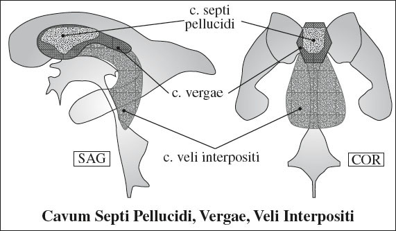
√ triangular-shaped CSF space / cyst in pineal region
› above tela choroidea of 3rd ventricle
› below columns of fornices
√ separated from cavum vergae by crura of fornices
VENTRICLES
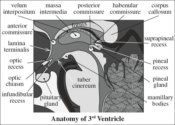
Third Ventricle
Position: in midline of diencephalon
Walls:
› inferiorly: curving floor of 3rd ventricle formed
» anteriorly by median eminence and tuber cinereum
» posteriorly by mammillary bodies + midbrain posterior to tuber cinereum
› superiorly: curving roof with 5 layers:
(1) body of fornix
(2) superior layer of tela choroidea
(3) vascular layer = velum interpositum containing bathed in CSF:
(a) internal cerebral veins
(b) medial posterior choroidal arteries
(4) inferior layer of tela choroidea
(5) choroid plexus of 3rd ventricle
› superior lateral wall: formed by medial aspects of thalami
› inferior lateral wall: formed by hypothalamus anteriorly + subthalamus posteriorly
› massa intermedia = band of gray matter as a communication between 2 thalami across midline
› anteriorly: lamina terminalis
» anterosuperior border: anterior commissure
» anteroinferior border: optic chiasm
› posteriorly: pineal gland
Recesses: 2 anterior + 2 posterior recesses
(1) optic recess superiorly
(2) infundibular recess inferiorly = pars cava infundibuli formed by funnel-shaped proximal hypothalamic infundibulum (pituitary stalk)
» median eminence at base of infundibulum = raised portion of hypothalamic gray matter
(3) pineal recess in deep aspect of pineal gland
» posterior commissure in inferior wall of pineal recess
» habenular commissure in superior wall of pineal recess
(4) suprapineal recess above habenular commissure and below splenium of corpus callosum
Tela choroidea ventriculi tertii
= ependymal + glial layers forming choroid plexus (= richly vascularized tela) that projects into 3rd ventricle
(a) upper layer attached to lower surface of fornix
(b) lower layer attached anteriorly to stria medullaris thalami + posteriorly to superior surface of the pineal body
[tela, Latin = web], [khorion, Greek = membrane enclosing fetus, afterbirth] [chorioeides, Greek = membrane-like] [plexus, Latin = braid, network]
Interventricular Foramen of Monro
= Y-shaped communication between 2 lateral ventricles and 3rd ventricle
Location: posterior to anterior columns of fornix
Aqueduct of Sylvius
= opening at posterior aspect of 3rd ventricle below posterior commissure
MEGACISTERNA MAGNA
= GIANT CISTERNA MAGNA = ENLARGED CISTERNA MAGNA
= large retrocerebellar CSF space with normal vermis + cerebellar hemispheres
Prevalence: 1% of all brains
May be associated with: infarction, inflammation, infection (esp. CMV), chromosomal abnormalities (esp. trisomy 18)
Size of normal cisterna magna:
3–8 mm measured in midsagittal plane from posterior lip of foramen magnum to caudal margin of inferior vermis
√ intact vermis + normal 4th ventricle
√ paired linear echoes that join + descend toward base of posterior fossa = dural folds = inferior attachment of tentorium
√ NO enlarged posterior fossa, NO mass effect on cerebellum
DDx: arachnoid cyst
PINEAL GLAND
= PINEAL BODY = EPIPHYSIS CEREBRI = “Third eye”
Development: diverticulum lined by ependyma at the most caudal portion of diencephalic roof of 3rd ventricle → area of ependymal thickening → evaginates into pinecone-shaped mass during 7th week GA
Function: secretion of melanin
1. Regulation of long-term biologic rhythm (eg, onset of puberty)
2. Regulation of short-term biologic rhythm (eg, diurnal / circadian) due to photoperiodic clues via accessory optic pathway
Histo:
(a) lobules of pineocytes with dendritic processes (= specialized neuronal cells related to retinal rods and cones) make up 95% of population
(b) astrocytes as neuroglial supporting cells = 5% of population
(c) fibrovascular stroma
(d) corpora arenacea = concentric calcifications appear in adolescence (in 40% of patients 17–29 years old)
[arenacea, Latin = sandy]
Location: midline
suspended from pineal stalk that is attached to upper aspect of posterior border of 3rd ventricle
(a) inferior to splenium + vein of Galen
(b) superior to tentorium + superior colliculi
(c) posterior to cistern of velum interpositum (= cistern of transverse fissure)
(d) surrounded by CSF of quadrigeminal cistern
Size: 10–14 mm
√ enhancement with contrast ← absence of blood-brain barrier
Related posts:


Stay updated, free articles. Join our Telegram channel

Full access? Get Clinical Tree


