Orbits


Orbits Axial 1


Orbits Axial 1


Orbits Axial 2


Orbits Axial 2


Orbits Axial 3


Normal Anatomy


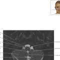
The lenses are normally biconvex in shape. If this configuration is replaced by a thin line, the patient likely has had the lens removed because of cataracts and replaced with an artificial lens.


Orbits Axial 3


Orbits Axial 4


Orbits Axial 4


Orbits Axial 5


Pathologic Process


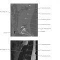
Note the thin superior ophthalmic vein (SOV). Dilatation should lead to consideration of increased pressure within the cavernous sinus, as in carotid cavernous fistula (CCF) following trauma. Early, asymmetric filling of the cavernous sinus in the arterial phase confirms the diagnosis of CCF (see also Orbits Coronal 1 ).

Only gold members can continue reading. Log In or Register to continue

Stay updated, free articles. Join our Telegram channel

Apr 4, 2019 | Posted by in NEUROLOGICAL IMAGING | Comments Off on Orbits

Full access? Get Clinical Tree

Get Clinical Tree app for offline access