Localization: Osteoblastoma shows evident predilection for the vertebral column (posterior arch) and the sacrum, but it may occur in any skeletal site.
Clinical: In the spine, it presents symptoms similar to osteoid osteoma (pain, scoliosis) with frequent signs of root compression. Usually it grows slowly but aggressive lesions manifest a rapid growth with severe symptoms due to the peritumoral inflammation.
Imaging: It is an osteolytic tumor containing a variable extent of osseous-type mineralization. Its size varies from 2 to 10 cm, the majority being between 3 and 5 cm. The tumor may be central, eccentric, and rarely periosteal. It tends to be roundish, with margins often demarcated by a rind of bone sclerosis, not as dense as in osteoid osteoma. The cortex may be destroyed with intense periosteal reaction. In aggressive lesions, the limits may appear blurred. Rarely the tumor blows the bone out or contains cystic spaces, similar to an ABC. A regional osteoporosis may be associated. The isotope bone scan is very hot. CT at best depicts intratumoral radio densities. MRI may show extensive peritumoral inflammatory reaction. Angiography reveals tumor vascularity.
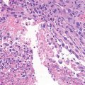
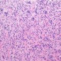
Histopathology: The tissue is compact, reddish brown, and of soft-to-gritty consistency. Occasionally wide cavities typical of ABC are observed. The cortex is thinned, expanded, and sometimes absent, with a pseudo-capsule covering the tumor.
Microscopically, the tumor consists of large osteoblasts producing an osteoid and woven bone. Trabeculae are usually thin, with a regular “organoid” pattern. Osteoblasts rim the trabeculae. Cytologic features of activity (large cytoplasm, plump dark nuclei, and evident nucleolus) may be present. Mitotic figures are rare and typical. Large cells with bizarre hyperchromatic nuclei may be seen; they are never in mitosis and are interpreted as regressive cells (the so-called pseudomalignant osteoblastoma). Intertrabecular tissue contains a loose fibrovascular stroma, with abundant capillaries. The interface between the tumor and surrounding bone is sharp with no permeative pattern (d. d. vs. osteosarcoma).
Course and Staging: Most osteoblastoma are actively growing but well contained (stage 2). Occasionally, they are more invasive, bulging into the soft tissues (stage 3). Rarely the tumor appears almost quiescent and heavily mineralized so that it can be approximated to a stage 1 lesion. The vast majority of “osteoblastomas” that end up metastasizing to the lungs, leading to patient demise, were probably osteoblastoma-like osteosarcomas from the beginning. It is very important to assess the matrix of the “osteoblastomatous” lesion with the host bone. If it permeates the marrow spaces and traps the host lamellar bone, the lesion is an osteosarcoma, osteoblastoma-like.
Related posts:


Stay updated, free articles. Join our Telegram channel

Full access? Get Clinical Tree


