Localization: It prevalently occurs in the appendicular skeleton, while it is rare in the trunk, with the exception of the spine (mostly localized in the posterior arch). In the limbs, it prevails in the proximal femur, but it is also common in the other long bones where it is more often diaphyseal or metadiaphyseal. It may occur also in the short bones, like talus. When occurring in long shafts, it involves the cortex, but also in cancellous areas, it is localized at the surface rather than in the center of the bone.
Clinical: The almost constant and often only symptom is pain, with a typical tendency to increase during the night, relieved by nonsteroidal anti-inflammatory drugs. When localized close to a joint, limited motion and chronic synovitis can be observed. In the spine, it may cause muscular spasm with stiff scoliosis.
Imaging: The basic radiographic element is a small (1–2 cm) rounded area of osteolysis (“nidus”), surrounded by a halo of bone sclerosis. The lesion is located at the center of this sclerosis that can be so thick that the small nidus cannot be seen except by CT. In cancellous bone areas, the halo of sclerosis may be less prominent. The isotope bone scan is constantly positive, showing a small, rounded area of intense uptake centered in a more diffused halo, thus resembling a “headlight in the fog.” CT always demonstrates the nidus and its localization, thus permitting an adequate surgical plan. Angiography may demonstrate the high vascularity of the nidus. MRI, instead, is of little value because it is inferior to CT in depicting the nidus and showing inflammation of the surrounding tissue; it may also confuse the diagnosis.
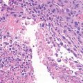
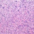
Histopathology: Small roundish and hyperemic tumor, softer than the surrounding bone. Microscopic view shows a packed mesh of thin, contorted woven bone trabeculae, with osteoblastic rimming, osteoclasts, and numerous dilated capillaries. The osteoid may tend to mature in the center of the nidus. This calcified center corresponds to the radio dense nucleus of the nidus. The surrounding host bone appears as a reactive bone, more or less mature and sclerotic. The soft tissues around the lesion show chronic inflammation.
Course and Staging: Left untreated, it increases very slowly. Actually, there are cases, managed by nonsteroidal anti-inflammatory drugs, where pain subsided after some years of treatment and eventually even the nidus regressed. The tumor can be staged 1 or 2 in all cases.
Treatment and Prognosis: Surgery has been historically the mainstay of treatment; accurate intralesional curettage has been the treatment of choice in most centers until the late 1990s. Nowadays CT-guided percutaneous radiofrequency or laser ablation is considered the treatment of choice. The success rate of this approach is usually more than 90 % based on pain relief. Surgery remains an option in cases refractory to percutaneous ablation.
Related posts:


Stay updated, free articles. Join our Telegram channel

Full access? Get Clinical Tree


