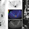
Fig. 27.1
A 13-year-old boy suffered a traumatic fracture of the left superior articular process of the fifth vertebra while playing football. Coronal (a–c), sagittal (d–f), and axial (g–i) CT with bone window (a, d, g), PET (b, e, h), and PET/CT fusion (c, f, i) images show focal FDG uptake corresponding to the fracture site

Fig. 27.2
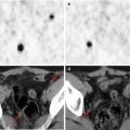
A 15-year-old boy underwent a PET evaluation during chemotherapy for Hodgkin’s lymphoma, stage III. Axial bone window CT (a), PET (b), and PET/CT fusion (c) images show 18F-FDG uptake in the left anterior iliac spine (yellow arrow in a). The diagnosis was a traumatic fracture subsequent to bone marrow biopsy
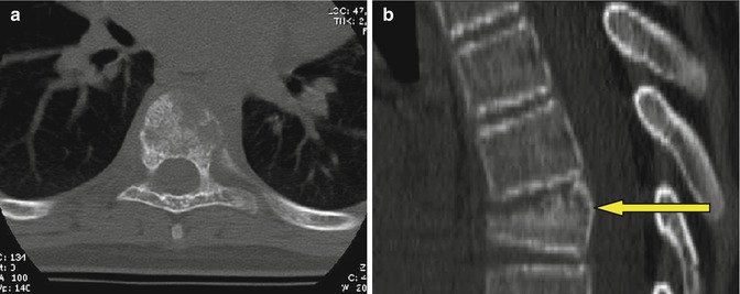
Fig. 27.3


An 11-year-old girl was admitted for dorsal pain, without trauma. Bone scintigraphy showed an accumulation in the fifth and sixth vertebrae. Axial (a) and sagittal (b) bone window CT shows a lytic lesion in the sixth dorsal vertebra (yellow arrow in b), with pathological findings in the left paravertebral soft tissues
Stay updated, free articles. Join our Telegram channel

Full access? Get Clinical Tree


