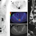
Fig. 7.1
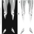
Coronal CT (a), PET/CT fusion (b), and axial CT and PET/CT fusion (c) images of the legs show focal 18F-FDG uptake in the upper right popliteal cavity, lateral to the femoral biceps muscle. A PET-guided biopsy proved the recurrence of acute myeloid leukemia
Case 2
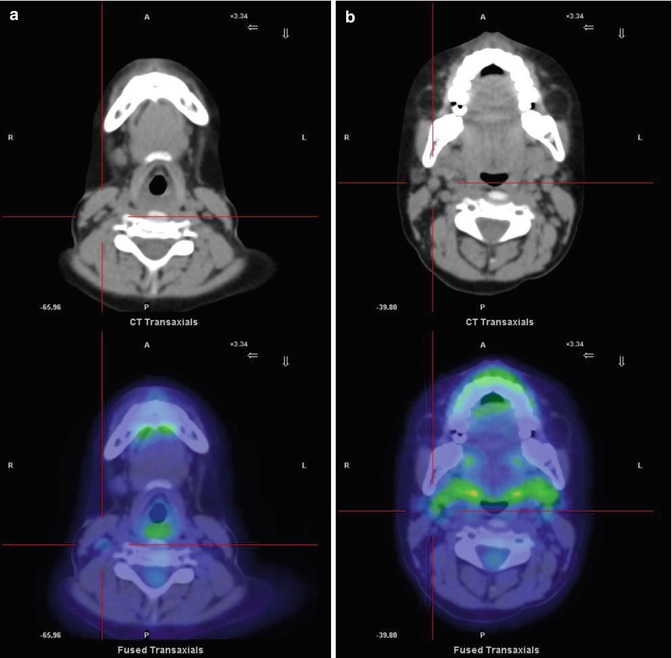
A 5-year-old boy was diagnosed and treated for pre-B ALL. Four years later, he was reevaluated for an isolated lymphadenopathy of the head–neck region, without signs of inflammation on the overlying skin. There was no serological evidence of a recent infection nor were there any notable changes after empirical antibiotic and anti-inflammatory therapy. The outcome is shown in Figs. 7.2 and 7.3.
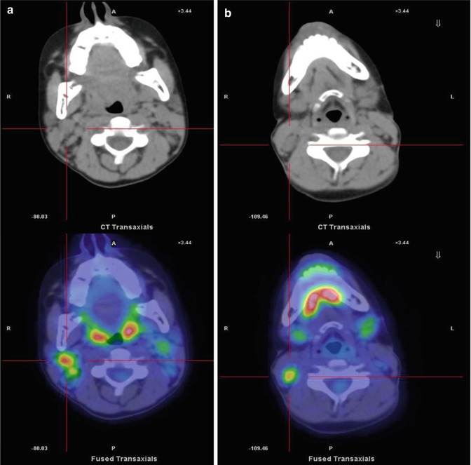

Fig. 7.2
Pretreatment PET/CT scan. (a, b) Axial CT and PET/CT fusion images show pathological 18F-FDG uptake by the right laterocervical lymph nodes
