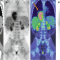
Fig. 18.1
A 12-year-old girl with a positive biopsy for malignant ovarian teratoma. Maximum intensity projection (a) shows diffuse pathological FDG uptakes in the pelvis and upper abdomen. Axial abdominal window CT and PET/CT fusion images (b) show the FDG-avid lesions in the pelvis
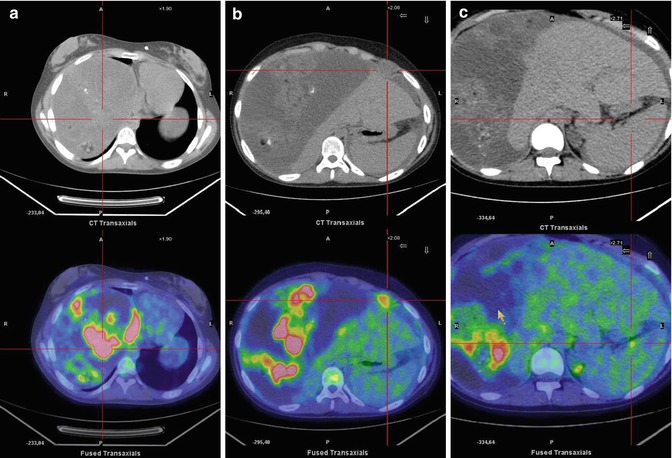
Fig. 18.2
Same patient. (a–c) Axial abdominal window CT and PET/CT fusion images show diffuse subglissonian (a), peritoneal (b), and peri-splenic (c) FDG-avid lesions
Teaching Point
Related posts:


Stay updated, free articles. Join our Telegram channel

Full access? Get Clinical Tree


