Paediatric oncology

Chapter 33 Paediatric oncology


Introduction


Cancer in childhood is uncommon. In the UK, approximately 1500 children below the age of 15 develop cancer each year. Approximately one individual child in 500 will develop cancer before the age of 15. The range of childhood cancers is very different from that seen in the adult population. Table 33.1 summarizes data on relative incidence of the various tumour types from the US Surveillance, Epidemiology and End Results (SEER) programme.


Table 33.1 Surveillance, epidemiology and end results (SEER) programme registrations 1975–2001, annual incidence rate per 1   000    000 and proportion of children aged 0–14 with cancer


Disease group Annual incidence rate per 1   000   000 Percentage of total
Acute lymphoblastic leukaemia (ALL) 32.8 23.6
Acute non-lymphoblastic leukaemia (ANNL) 6.4 4.6
Other leukaemias 3.0 2.2
All leukaemias 42.2 30.3
Astrocytoma 14.5 10.4
Medulloblastoma/PNET 6.6 4.7
Ependymoma 2.7 1.9
Intracranial germ cell tumours 1.1 0.8
Other CNS tumours 5.7 4.1
All CNS tumours 29.4 21.1
Hodgkin’s disease 6.0 4.3
Non-Hodgkin’s lymphoma 7.8 5.6
Osteosarcoma 3.5 2.5
Ewing’s sarcoma/peripheral PNET 2.4 1.7
Rhabdomyosarcoma 4.8 3.5
Other sarcoma 5.3 3.8
Neuroblastoma 10.3 7.4
Wilms’ tumour 8.3 6.0
Total 139.1  

The evolution of the multidisciplinary care of children with cancer has been one of the success stories of modern oncology. Paediatric oncology collaborative groups have been very successful at entering a high proportion of children into clinical trials. In North America, clinical research has been coordinated via the Paediatric Oncology Group (POG), Children’s Cancer Group (CCG), Intergroup Rhabdomyosarcoma Study Group (IRSG) and National Wilms’ Tumour Study Group (NWTS). In 1999, these were amalgamated to form the Children’s Oncology Group (COG), the largest paediatric oncology collaborative group in the world.


In the UK, treatment is coordinated by the network of twenty-two Children’s Cancer and Leukaemia Group (CCLG) (formerly UKCCSG) paediatric oncology centres. Increasingly, collaboration is across European boundaries, with clinical trials coordinated via the International Society of Paediatric Oncology (SIOP).


Currently, approximately two-thirds of children treated for cancer can expect to be long-term survivors. For most diseases, this has been brought about largely as a result of the incorporation of chemotherapy as part of a multimodality approach, including surgery and radiotherapy. Since the introduction of chemotherapy into treatment programmes in the 1960s and 1970s, the proportion of children surviving cancer has shown a gratifying increase.


Approximately 40–50% of children with cancer receive radiotherapy as part of their initial treatment. Radiotherapy is an important modality of therapy in local tumour control and the majority of paediatric tumours are radiosensitive. Cure, however, often comes ‘with a cost’ as a result of the long-term sequelae of treatment. Long-term effects of radiotherapy include impaired bone and soft tissue growth, impaired neuropsychological development as a result of irradiation of the central nervous system (CNS) and radiation-induced malignancy. Increasing awareness of long-term effects in the 1970s and 1980s led to a general decline in the use of radiotherapy. However, more recently, it has become evident that chemotherapy is also associated with long-term side effects, including late myocardial damage due to anthracyclines, nephrotoxicity due to cisplatin or ifosfamide, and secondary leukaemia related to a number of chemotherapeutic agents including alkylating agents. The overall aim of paediatric oncology programmes is to maximize the chance of cure with the minimum impact of likely long-term effects of treatment. Continued vigilance for long-term effects of treatment is essential. This is ideally performed in the setting of dedicated long-term follow-up clinics and employing national treatment-related guidelines for long-term follow up, such as those produced by the CCLG and the Scottish Intercollegiate Group.


It is very important that the administration of chemotherapy and radiotherapy for children should be undertaken only in specialized oncology centres treating relatively large numbers of children. In the UK, these centres are usually affiliated to the CCLG. The multiprofessional paediatric radiotherapy team should include a specialist paediatric therapy radiographer, specialist nurse and play specialist. Young children, particularly those under the age of three to four, find it very difficult to lie still for radiotherapy planning and delivery, especially when a Perspex head shell is required. Sedation sufficient to ensure immobilization is difficult to achieve without it persisting for several hours and it is not feasible for this to be administered daily. Because of the importance of immobilization, short-acting general anaesthesia, such as DiprivanÒ (propofol), is sometimes needed. The daily fasting for this results in surprisingly little disruption to nutrition. An experienced play therapist can be very helpful in preparing the child for radiotherapy and may avoid the need for daily anaesthesia for some children.


Toxicity of radiotherapy for children


Subacute effects


Long-term effects


Central nervous system


Paediatric radiation oncology involves a significant amount of time devoted to the treatment of children with brain tumours, and consideration needs to be given to the toxicity of therapy.


Radionecrosis is rare below 60   Gy, and generally occurs with a latency of 6 months to 2 years. It results from a direct effect on glial tissue. It is very unusual to have to deliver a dose of 60   Gy to any part of the CNS for a child and, for the radical treatment of children with brain tumours, it is very uncommon to exceed a dose of 50–54   Gy. It occurs in approximately 50% of patients treated by interstitial implantation for recurrent brain tumours following prior radical external beam radiotherapy. The clinical effects of radionecrosis vary according to the site within the CNS and are most devastating in the spinal cord. Radionecrosis of the spinal cord in children has been seen as a consequence of the interaction between radiation and cytosine arabinoside given intrathecally for metastatic rhabdomyosarcoma.


Necrotizing leukoencephalopathy may be seen when cranial irradiation is followed by high dose methotrexate for the treatment of leukaemia. The clinical features include ataxia, lethargy, epilepsy, spasticity and paresis.


Neuropsychological effects. The effects of cranial radiotherapy are now well established. Data from children treated with prophylactic radiotherapy for leukaemia have demonstrated that when compared with siblings, children given 24   Gy prophylactic cranial irradiation show an approximate fall in IQ of 12 points. Following higher radiation doses given for brain tumours, an increased risk of learning and behaviour difficulties is seen. An important risk factor for the incidence and severity of neuropsychological long-term effects is the age at diagnosis. Other factors include the impact of direct and indirect tumour-related parameters, treatment parameters with neuropsychological long-term effects worse for whole brain compared with partial brain irradiation, concomitant use of some chemotherapeutic agents, premorbid patient characteristics, such as intelligence, and the quality of ‘catch up’ education.


Chemotherapy/radiotherapy interactions


Interactions between radiation and chemotherapy are complex and poorly understood. Interactions can be exploited in order to attempt to improve disease-free survival. The most frequently employed mechanism in paediatric oncology is ‘spatial cooperation’ whereby chemotherapy and radiotherapy are combined to exploit their differing roles in different anatomical sites. Examples are the use of radiation for local control of a primary, with chemotherapy for subclinical metastatic disease such as in the treatment of Ewing’s sarcoma.


Chemotherapy and radiotherapy may be combined with the aim of increasing tumour cell kill without excess toxicity. An example is the use of combined chemotherapy and radiotherapy for children with Hodgkin’s disease. It may be possible to reduce the intensity of both treatment modalities and hopefully reduce long-term morbidity. When using combined modality therapy the aim is to improve the therapeutic ratio. Many protocols for children involve the use of concurrent chemotherapy and radiotherapy. It is essential to be vigilant for additional early or long-term morbidity. Clinically important chemotherapy/radiotherapy interactions are often unpredictable and their mechanisms poorly understood. Actinomycin-D and cisplatin increase the slope of the radiation dose–response curve and actinomycin-D inhibits the repair of sublethal damage (SLD). Clinical interactions include enhanced skin and mucosal toxicity when radiation is followed by actinomycin-D (the ‘recall phenomenon’), enhanced bladder toxicity when chemotherapy is combined with cyclophosphamide, enhanced CNS toxicity from combined radiation and methotrexate, cytosine arabinoside or busulfan, and the enhanced marrow toxicity from wide field irradiation and many myelotoxic chemotherapeutic agents. In the case of the effect of combined radiation and anthracyclines, such as doxorubicin on the heart, doxorubicin has its effects on the myocytes and radiation on the vasculature.


Radiotherapy quality assurance (QA)


Because of the high cure rate for most childhood cancers, it is important to achieve local tumour control and avoid a ‘geographical miss’. It is also important to avoid unnecessarily large field sizes, in order to minimize long-term effects.


It is essential for all radiotherapy departments to deliver the highest possible standard of radiotherapy for all patients including children. Many radiotherapy departments have adopted quality systems.


In a number of studies, particularly those employing craniospinal radiotherapy for medulloblastoma, the accuracy of delivery of radiotherapy has impacted upon tumour control and patient survival. In many North American paediatric multicentre studies, radiotherapy quality, including beam data, dose prescription, planning and verification films are reviewed centrally in the Quality Assurance Review Centre (QARC) situated in Providence, Rhode Island. In the majority of European studies, QA is not as well organized. Ideally, review of radiotherapy simulator or verification films by study coordinators should be sufficiently fast to provide feedback early in the course of radiotherapy so that the treatment plan can be modified if necessary. The electronic transmission of planning films and associated clinical data can facilitate this process. This is logistically difficult but has been achieved in the USA for radiotherapy administered to children treated within POG, CCG and more recently COG trials. In Europe, where funding for these activities is more problematic, this has been achieved in Germany for radiotherapy for Ewing’s sarcoma.


Leukaemia


The improvement in survival of children with acute lymphoblastic leukaemia (ALL) was one of the early successes of paediatric oncology. Currently, more than 70% are long-term survivors. The leukaemias account for the most frequent group of paediatric malignancies. Approximately 80% have ALL and 20% acute non-lymphoblastic leukaemia (ANLL), usually acute myeloid leukaemia (AML) or rarely chronic myeloid leukaemia (CML).


Current treatment for ALL is stratified according to risk status based on presenting white count and cytogenetic profile. The four phases of treatment are:


During the 1960s and 1970s, the routine use of prophylactic whole brain radiotherapy and intrathecal methotrexate reduced the risk of CNS relapse to less than 10%. Whole brain radiotherapy may be employed for patients who present with CNS involvement (Figure evolve 33.1 image). Patients are immobilized in a head shell and treated with lateral-opposed 4–6   MV megavoltage fields which may be centred on outer canthus to minimize divergence into the contralateral lens. Shielding will cover the face, dentition, nasal structures and lenses. The clinical target volume (CTV) includes the intracranial meninges extending inferiorly to the lower border of the second or third cervical vertebra. Great care is taken to include the cribriform fossa, temporal lobe and base of skull. Although the lens is shielded, as much of the posterior orbit as possible is included as ocular relapses occasionally occur. The CTV is localized with lateral simulator radiographs or, more recently, computed tomography (CT) simulation. The prescribed dose in current UK protocols is 24   Gy in 15 fractions of 1.6   Gy daily.


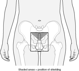
Boys who suffer a testicular relapse are treated with testicular radiotherapy (Figure 33.2). The technique employed is an anterior field, generally electrons or orthovoltage (200–300   kV). The CTV includes both testes, scrotum and inguinal canal superlaterally as far as the deep inguinal ring with shielding of non-target skin and perineum. The prescribed dose is 24   Gy in 12 fractions of 2.0   Gy daily.


As for adults, children with ANNL are treated with intensive multidrug chemotherapy, which can achieve a survival rate of 60%. Bone marrow transplantation (BMT) is frequently employed for children who have an HLA-matched sibling. A survival rate of 65% can be achieved for children in first complete remission treated with BMT as consolidation therapy for acute myeloid leukaemia.


Total body irradiation (TBI)


TBI is an important technique used together usually with high dose cyclophosphamide (Cyclo-TBI) as the conditioning regimen prior to BMT for adults and children. Bone marrow donors for BMT are generally HLA-matched siblings. However, increasingly, volunteer unrelated donors from donor panels donate marrow, resulting in a significant increase in the number of patients for whom BMT can be considered.


Techniques for TBI have evolved in different departments, generally depending on availability of treatment facilities. Modern linear accelerator design and field sizes allow the use of large anterior and posterior fields. TBI dosimetry is usually based on in vivo measurements.


As an example of a TBI technique, the Leeds technique is described. The patient lies in the lateral position in an evacuated polystyrene immobilization bag. Hands are placed under chin to provide ‘lung compensation’ (Figure evolve 33.3 image). Dosimetry is determined using in-vivo measurements performed at a ‘test dose’ of 0.2   Gy for each field. For such a large and complex target volume, it is not feasible to adhere to the ICRU 50 guidelines of a range of −5% to +7%, and a range of −10% to +10% is more realistic. The standard TBI dose for children in the UK is 14.4   Gy in eight fractions of 1.8   Gy twice daily with a minimum interfraction interval of 6 hours.

Only gold members can continue reading. Log In or Register to continue

Stay updated, free articles. Join our Telegram channel

Mar 7, 2016 | Posted by in GENERAL RADIOLOGY | Comments Off on Paediatric oncology

Full access? Get Clinical Tree

Get Clinical Tree app for offline access