and Filiz Özülker1
(1)
Nuclear Medicine, Okmeydani Training and Research Hospital, Istanbul, Turkey
11.1 Case 1: Pancreas Adenocarcinoma
History
A 73.-year-old female underwent tru-cut biopsy from mass lesion at liver and histopathology revealed metastasis of adenocarcinoma. A 18F-FDG PET/CT scan was performed in order to determine the primary malignancy.
Findings
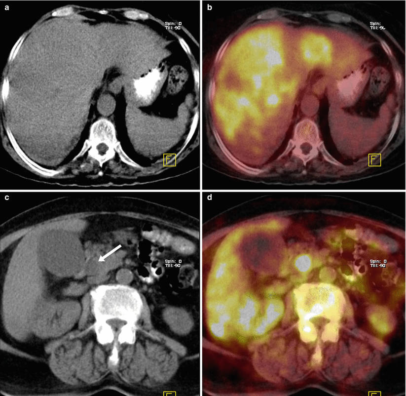
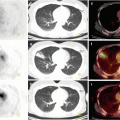
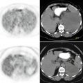
Fig. 11.1
Axial CT and fusion images show multiple hypermetabolic mass lesions at both lobes of liver (SUVmax 10) (a, b) and a focal hypermetabolic lesion at the head of the pancreas (arrow) (SUVmax 8.7) (c, d)
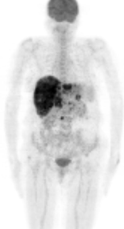
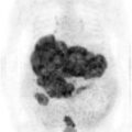
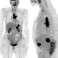
Fig. 11.2
MIP image
Related posts:
Stay updated, free articles. Join our Telegram channel

Full access? Get Clinical Tree


