CT: Linear high-density foci within dilated biliary tree
 US: Tubular echogenic structure, 3-6 mm in diameter, with central anechoic area (digestive tract of worm)
• Clonorchiasis: Flukes typically involve peripheral intrahepatic ducts, not GB, CBD, or PD
 CT/MR: Preferential dilatation of small peripheral intrahepatic ducts with intraductal high-density foci
CT/MR: Preferential dilatation of small peripheral intrahepatic ducts with intraductal high-density foci
• Fascioliasis: Flukes usually involve large intrahepatic ducts, extrahepatic duct, and GB (after liver involvement)

• Echinococcosis: Communication of hepatic hydatid cyst with small biliary radicles or rupture of cyst into bile ducts
 CT: High-attenuation material within dilated bile ducts, often contiguous with wall defect in hepatic hydatid cyst
CT: High-attenuation material within dilated bile ducts, often contiguous with wall defect in hepatic hydatid cyst 
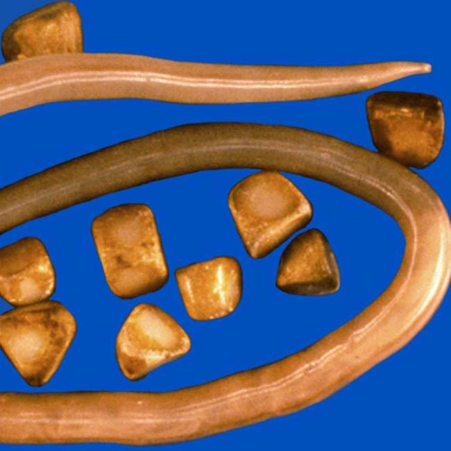
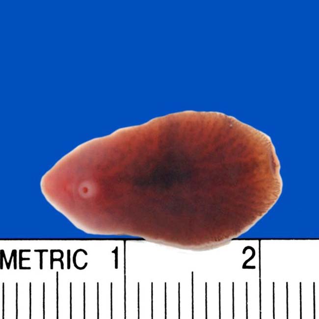
 from a biliary Ascaris worm.
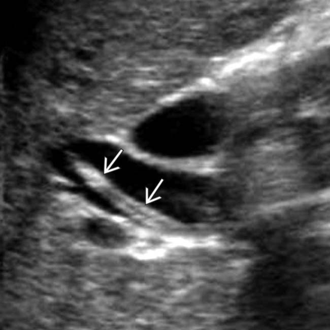
 . The worm was mobile at real-time examination. Central hypoechogenicity is thought to represent the digestive tract of the worm. (Courtesy A. Dasyam, MD.)
IMAGING
General Features
• Best diagnostic clue
 Echinococcosis: Dilated, debris-filled biliary ducts adjacent to ruptured hepatic hydatid cyst on CT, US, MR
 Echinococcosis: Dilated, debris-filled biliary ducts adjacent to ruptured hepatic hydatid cyst on CT, US, MR
• Location
 Clonorchiasis typically involves peripheral intrahepatic ducts, not gallbladder (GB), common bile duct (CBD), or PD (except in heavy infections)
 Clonorchiasis typically involves peripheral intrahepatic ducts, not gallbladder (GB), common bile duct (CBD), or PD (except in heavy infections)


CT Findings
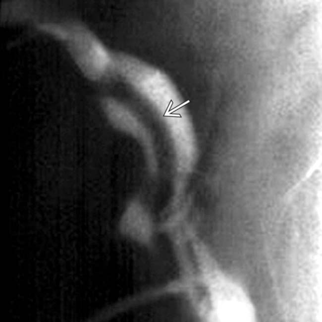
• Ascariasis/clonorchiasis: Intraductal high-density foci within dilated biliary tree due to biliary worms/flukes or debris
 Imaging evidence of complications, including peripancreatic inflammation due to pancreatitis, intrahepatic abscess, and abnormal biliary tree wall enhancement or heterogeneous parenchymal enhancement due to cholangitis
Imaging evidence of complications, including peripancreatic inflammation due to pancreatitis, intrahepatic abscess, and abnormal biliary tree wall enhancement or heterogeneous parenchymal enhancement due to cholangitis
• Echinococcosis: High-attenuation material within dilated bile ducts, often continuous with wall defect in hepatic hydatid cyst


MR Findings

• Clonorchiasis: Preferential peripheral biliary dilatation with low-signal filling defects on T2WI or MRCP
• Echinococcosis: Low-signal linear or rounded filling defects within dilated ducts, often with adjacent deformed hydatid cyst


Ultrasonographic Findings
• Grayscale ultrasound
 Ascariasis: Ultrasound very sensitive for worms in biliary system, but insensitive for worms in duodenum or ampulla (sensitivity for pancreatobiliary ascariasis only 50%)
 Clonorchiasis: Flukes appear as echogenic filling defects (without shadowing) in bile ducts which float with changes in position


Ascariasis: Ultrasound very sensitive for worms in biliary system, but insensitive for worms in duodenum or ampulla (sensitivity for pancreatobiliary ascariasis only 50%)– Tubular echogenic structure, 3-6 mm in diameter, with central anechoic area (digestive tract of worm)
Clonorchiasis: Flukes appear as echogenic filling defects (without shadowing) in bile ducts which float with changes in position– Often associated with bile duct stones (including hepatolithiasis), which are echogenic and demonstrate posterior acoustic shadowing
Related posts:

Stay updated, free articles. Join our Telegram channel
Full access? Get Clinical Tree
