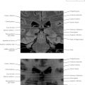
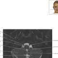
Paranasal Sinuses Axial 2
Normal Anatomy
The sphenoid sinuses are at the center of the skull base, immediately below the sella turcica (see Chapter 7 ). In ancient Egypt, mummification would involve driving a metal hook through the nasal passages and the sphenoid sinuses into the brain, which would be removed through the nostrils. This tract is used in modern days to pass a scope into the sella turcica to remove pituitary adenomas.

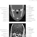
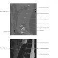
Paranasal Sinuses Axial 3
Diagnostic Consideration
In the setting of trauma, careful evaluation of CT bone windows is important to identify fractures, particularly around critical structures such as the carotid canal. On MRI, subtle bone fractures may not be directly visible; therefore, secondary signs, such as the presence of a hemorrhagic fluid level in the paranasal sinuses may be the only indication of traumatic injury.


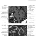
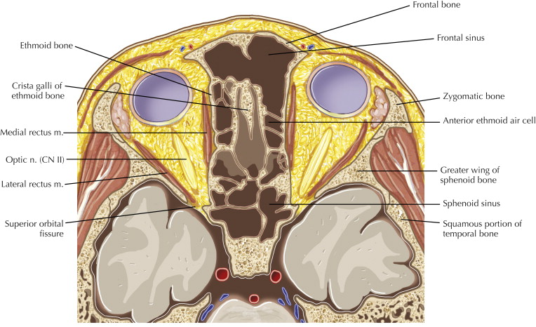
Paranasal Sinuses Axial 5

Normal Anatomy
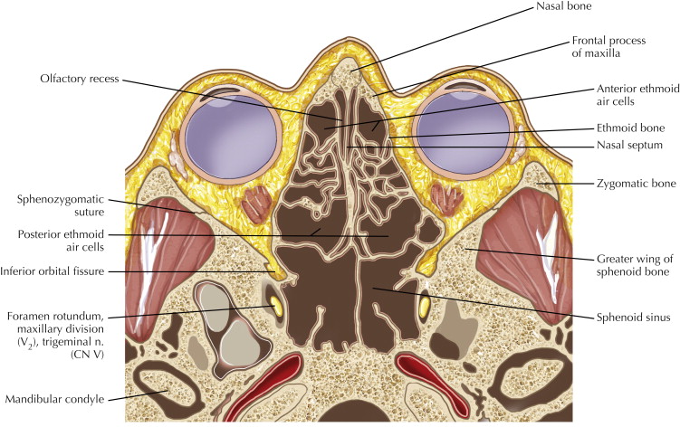
The pterygopalatine fossa houses the second division of the fifth cranial nerve (CN V), the maxillary nerve (V 2 ). The ganglion associated with V 2 lying in this fossa is known as Meckel’s (pterygopalatine) ganglion, whereas the Gasserian (trigeminal) ganglion lies in Meckel’s cave. Loss of the fat surrounding the nerve and ganglion is seen in perineural infiltration by malignancy.

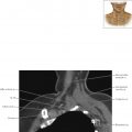
Paranasal Sinuses Axial 6