Part 9 Infection and Inflammation
Case 104

Clinical History
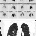
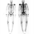
27-year-old male with end-stage renal disease secondary to focal segmental glomerulonephritis presented with fevers and was found to have methicillin-susceptible Staphylococcus aureus bacteremia without known source (▶Fig. 104.1).
Key Finding
Focal abnormal uptake on tagged white blood cell (WBC) scintigraphy
Top 3 Differential Diagnoses
Infection. WBCs can be removed from the patient and tagged with either In-111 oxine or technetium-99m (Tc-99m) hexamethylpropyleneamine oxime (HMPAO) for use with whole-body imaging. Tagging can be done in vivo, in vitro, or with a mixed technique. The in vitro technique is most commonly used as it is most efficient. In-111 oxine has the advantage of a long half-life and no hepatobiliary, genitourinary, or intestinal clearance. Tc-99m HMPAO provides better imaging statistics and less radiation dose to the patient. Both are useful for many indications, many of which have been replaced by CT and magnetic resonance imaging (MRI). In-111 oxine WBC imaging is most useful for renal infection, chronic osteomyelitis, and fever of unknown origin (FUO). Tc-99m HMPAO WBC imaging is most useful for acute osteomyelitis of the appendicular skeleton, vascular graft infection, and soft-tissue infection. Both can be used for inflammatory bowel disease (IBD), with some studies showing improved sensitivity with early imaging with Tc-99m HMPAO, before hepatobiliary clearance interferes, due to the improved imaging statistics. Mucosal uptake may be sloughed limiting the utility of In-111 oxine. Abnormal uptake of either one of the tagged WBC imaging agents results from a combination of hyperemia, vascular permeability, and chemotaxis. Focal uptake is worrisome for pyogenic infection. Infectious uptake is generally as intense as physiologic liver uptake, with more intense uptake raising suspicion for abscess. Single-photon emission computed tomography (SPECT)/CT imaging can provide additional characterization.
Inflammation. As WBC uptake is not the only uptake mechanism for radiolabeled WBCs, increased uptake is not specific for pyogenic infection. Any inflammation can result in hyperemia and increased vascular permeability, and therefore, result in increased uptake of either In-111 oxine WBC or Tc-99m HMPAO WBC. In general, inflammatory uptake may be less intense than infectious uptake, more along the level of physiologic bone marrow activity.
Healing. Both postsurgical and traumatic healing will demonstrate increased radiolabeled WBC activity. Correlation with clinical history is important.
Additional Diagnostic Consideration
Bleed: While radiolabeled WBC distribute in the reticuloendothelial system (spleen, liver, and bone marrow), they also circulate within the blood pool. It is therefore possible for abnormal activity to be seen with active bleeding. Gastrointestinal (GI) bleeding has been seen on tagged WBC imaging.
Diagnosis
Left upper arm arteriovenous graft hematoma.
✓ Pearls
Tagged WBC uptake can be seen with infection, inflammation, healing, and bleeding.
As a general guideline, uptake in abscess may be greater than infection and inflammation.
Fluorine-18 fluorodeoxyglucose positron-emission tomography (F-18 FDG PET) imaging has proved equally or more useful than tagged WBC imaging for many indications.
Suggested Readings
Becker W, Meller J. The role of nuclear medicine in infection and inflammation. Lancet Infect Dis. 2001; 1(5):326–333 Cortés J, Villaverde J, Garrido M, Pombo M, Ruibal A. Incidental detection of gastrointestinal bleeding from an aortoenteric fistula on 99 mTc leukocyte scintigraphy. Clin Nucl Med. 2012; 37(11):1123–1125 Gotthardt M, Bleeker-Rovers CP, Boerman OC, Oyen WJ. Imaging of inflammation by PET, conventional scintigraphy, and other imaging techniques. J Nucl Med Technol. 2013; 41(3):157–169Case 105

Clinical History
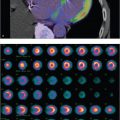
48-year-old female with sarcoidosis, lung and lymph node involvement (▶Fig. 105.1).
Key Finding
Increased breast uptake on Ga-67 citrate scintigraphy
Top 3 Differential Diagnoses
Hormone stimulation. Both prolactin and estrogen can stimulate breast uptake of Ga-67 citrate. Hyperprolactinemia results in intense, symmetric, increased bilateral breast uptake; most intense when seen in the immediate postpartum state. There may be relative photopenia in the subareolar region resulting in a “doughnut” configuration, likely due to peripheral lobular development stimulated by prolactin. The “doughnut” pattern can help differentiate from estrogen therapy which generally results in a more subareolar or diffuse pattern of uptake. Hyperprolactinemia can be seen with renal failure as well as a side effect of several medications, in addition to normal postpartum lactation.
Infection. Ga-67 citrate is primarily used as an infection/inflammation imaging agent due to its avidity for bacterial infections as well as acute and chronic inflammatory pathology. Asymmetric increased breast activity can be seen with mastitis. More focal uptake may suggest an abscess.
Lymphoma. Ga-67 citrate has been used extensively for lymphoma evaluation, most prominently to help assess response to therapy, but now it has largely been replaced by fluorine-18 fluorodeoxyglucose positron-emission tomography (F-18 FDG PET) imaging. However, Ga-67 citrate scintigraphy does have good sensitivity for extranodal lymphoma. Secondary breast involvement is more common than primary breast lymphoma.
Additional Diagnostic Consideration
Primary breast pathology: Ga-67 citrate scintigraphy is not routinely used for evaluation of breast pathology, as uptake in primary breast malignancies is variable. However, Ga-67 citrate uptake has been seen in primary breast pathology, including invasive ductal carcinoma, fibroadenoma, and fibrocystic change.
Diagnosis
Physiologic breast uptake.
✓ Pearls
Symmetric increased Ga-67 citrate breast uptake is almost always due to hormonal stimulation.
Symmetric increased breast uptake may be seen in renal failure due to associated hyperprolactinemia.
Asymmetric or focal Ga-67 citrate breast uptake is more concerning, but not always pathologic.
Suggested Readings
Kim YC, Brown ML, Thrall JH. Scintigraphic patterns of gallium-67 uptake in the breast. Radiology. 1977; 124(1):169–175 Moralidis E, Mandala E, Venizelos I, et al. A breast fibroadenoma mimicking an extranodal deposit of Hodgkin’s lymphoma in 67Ga imaging. Br J Radiol. 2009; 82(975):e58–e62 Taher MA, Navalkissoor S, Hilson AJ, Buscombe JR. Gallium citrate uptake is a marker of breast malignancy: true or false? Nucl Med Rev Cent East Eur. 2007; 10(1):21–22Case 106

Clinical History
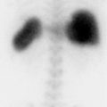
73-year-old female referred for left knee pain 5 years after total knee arthroplasty, concern for infection and/or loosening (▶Fig. 106.1).
Key Finding
Genitourinary tract uptake on In-111-labeled WBC scan
Top 3 Differential Diagnoses
Infection. In-111 is cyclotron produced, decays by electron capture releasing 173 and 247 keV gamma photons, with a physical half-life of 67 hours. In-111 oxine-labeled WBC scintigraphy is frequently used for infection imaging. Labeling is most commonly done in vitro which allows for the best efficiency and results in irreversible binding of the radionuclide to intracellular components primarily within neutrophils. Uptake mechanism is via chemotaxis of WBC. Imaging is generally performed around 24 hours after injection of the radiolabeled WBC. Normal distribution entails higher uptake within the spleen than liver and bone marrow. There is no hepatobiliary, intestinal, or genitourinary clearance. It is important to remember that there is no normal physiologic renal activity seen with In-111 WBC, since renal activity is so frequently seen on other nuclear medicine exams, including Tc-99m hexamethylpropyleneamine (HMPAO)-labeled WBC which has both hepatobiliary and genitourinary clearance. Urinary tract infection may present as activity within the genitourinary collecting system on In-111 WBC scintigraphy. Parenchymal renal uptake suggests pyelonephritis.
Transplant complications. In-111 WBC uptake in a transplant kidney is not specific. Uptake can be seen with rejection, acute tubular necrosis (ATN), and infection. Immunosuppressive therapy may limit the effectiveness of tagged WBC scintigraphy.
Other radiopharmaceutical. In-111 WBC scintigraphy is often performed in conjunction with other nuclear medicine exams, such as Tc-99m medronic acid (MDP) bone scan and Tc-99m SC bone marrow imaging for osteomyelitis evaluation. Genitourinary uptake, in this setting, may be due to normal renal clearance of one of the Tc-99m based radiopharmaceuticals.
Diagnosis
Urinary tract infection.
✓ Pearls
In-111-tagged leukocytes demonstrate no hepatobiliary, intestinal, or genitourinary clearance.
Genitourinary uptake on an In-111 WBC scan is abnormal and concerning for infection.
In-111 WBC uptake in a renal transplant is not specific and can be seen with ATN, rejection, and infection.
Suggested Readings
Hughes DK. Nuclear medicine and infection detection: the relative effectiveness of imaging with 111 In-oxine-, 99 mTc-HMPAO-, and 99 mTc-stannous fluoride colloid-labeled leukocytes and with 67Ga-citrate. J Nucl Med Technol. 2003; 31(4):196–201, quiz 203–204 Love C, Palestro CJ. Radionuclide imaging of infection. J Nucl Med Technol. 2004; 32(2):47–57, quiz 58–59Case 107

Clinical History
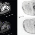
63-year-old female with fever, elevated inflammatory markers, and low back with new neurologic deficits, concern for epidural abscess. Too obese for MRI (▶Fig. 107.1).
Key Finding
Abnormal intra-abdominal uptake on tagged leukocyte scintigraphy
Top 3 Differential Diagnoses
Infection. Either In-111 oxine or Tc-99m hexamethylpropyleneamine (HMPAO)-tagged WBC scintigraphy remains useful tool for localization of infection. Sensitivity for abdominal abscess is greater than both ultrasound (US) and CT, although CT does have higher specificity. Leukocyte scintigraphy generally involves 24-hour imaging, making it less useful if immediate diagnosis is needed. Tagged WBC scan is most useful in a patient with presumed abdominal infection without localizing signs and negative anatomic imaging. Utilizing single-photon emission computed tomography (SPECT)/CT imaging allows for immediate anatomic localization and procedure planning, if the associated anatomic imaging is of high enough quality. In-111 oxine WBC scan is preferred for abdominal infection imaging as there is no hepatobiliary, intestinal, or genitourinary clearance.
Inflammation. One of the main limitations of leukocyte scintigraphy is difficulty in distinguishing infection from inflammation. Although as a general rule, infection will have uptake equal to or greater than the liver, and inflammation less than the liver, these patterns are not 100% specific. The uptake mechanism in inflammation includes hyperemia and vascular permeability, and less so WBC chemotaxis. Leukocyte scintigraphy can be useful for imaging IBD, as it is noninvasive, allows for imaging of the entire bowel, may distinguish between Crohn’s disease and ulcerative colitis, and does not require bowel preparation. While In-111 oxine WBC is generally preferred for intra-abdominal imaging, Tc-99m HMPAO WBC is preferred for IBD. This is due to early imaging and improved statistics, as late imaging with In-111 oxine WBC may be falsely negative due to sloughing mucosa.
Accessory spleen. Physiologic distribution of labeled leukocytes involves the reticuloendothelial system, more uptake in spleen than liver and bone marrow. Focal uptake in an accessory spleen in a patient with prior splenectomy can be confusing and mimic intra-abdominal infection. Correlation with anatomic imaging, or a Tc-99m SC liver spleen scan, can be confirmatory.
Related posts:

Stay updated, free articles. Join our Telegram channel

Full access? Get Clinical Tree


