May be some variant of B1 or B2, up to total gastrectomy
• Complications include recurrent tumor and acute or chronic sequelae of surgery
• Recurrent or new carcinoma
Local, lymph node, peritoneal, hematogenous
• Bezoar formation
Conforms to shape of stomach, traps air within
• Anastomotic leak
CT may detect indirect signs of leaks missed on upper gastrointestinal (GI) series (up to 50% of cases)
• Duodenal stump leakage
Loculated collection of fluid in subhepatic space
Rarely diagnosed on upper GI
• Jejunogastric intussusception
Rare complication of B2 procedure
• Afferent loop syndrome
Obstruction of afferent loop at or near anastomosis → dilation of duodenum
DIAGNOSTIC CHECKLIST
• Upper GI series is 1st-line test for detecting mechanical complications of gastric surgery
• CT is optimal test for general surveillance for postoperative complications
• PET/CT is optimal imaging test for surveillance of recurrent gastric carcinoma
• Abscessogram may identify leak as source of infection
(Left) Graphic depicts an isoperistaltic Billroth 2 gastrojejunostomy. The afferent limb , composed of the duodenum and a variable length of jejunum, carries pancreaticobiliary secretions toward the stomach, while the efferent limb carries fluid and food downstream.
(Right) Graphic depicts an antiperistaltic Billroth 2 procedure, in which the afferent loop enters the anastomosis from a left-to-right direction. This procedure is intended to reduce the prevalence of bile gastritis.
(Left) Film from a small bowel follow-through (SBFT) shows evidence of a prior Billroth 2 procedure and complete obstruction of antegrade flow of barium in the mid jejunum . At surgery, a phytobezoar was removed, which corresponded to the shape and size of the gastric remnant.
(Right) Film from an upper GI series shows evidence of a prior Billroth 1 procedure, along with persistent filling defects within the stomach that conform to the shape of the stomach, a bezoar.
IMAGING
General Features
• Many to most fluoroscopic exams of esophagus, stomach, and duodenum are now performed for patients who have surgically altered anatomy
• Some procedures are so common they are discussed separately
Postoperative state, esophagus
– Includes esophagectomy with gastric pull-through
Fundoplication complications
Bariatric surgery
• Goal for evaluating remaining procedures
Define expected postoperative anatomy
Describe imaging approaches to evaluation of postoperative patients
Describe imaging and clinical findings for various complications
Surgical Procedures
• Billroth 1 (B1) procedure
Antrectomy with gastroduodenostomy
Polya variation: Entire excised end of gastric stump is used for anastomosis
Hofmeister: Only a portion (usually greater curvature portion) is used
• Billroth 2 (B2) procedure
Distal gastrectomy with gastrojejunostomy
– Stomach may be anastomosed to Roux limb or loop of jejunum
– Anastomosis is side to side
– Variable length of duodenum and jejunum forms proximal or afferent loop


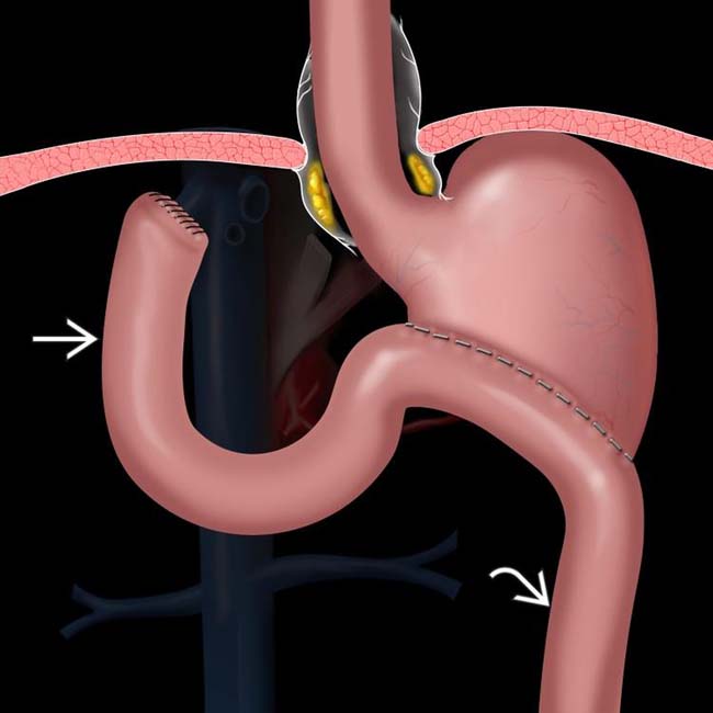
 , composed of the duodenum and a variable length of jejunum, carries pancreaticobiliary secretions toward the stomach, while the efferent limb
  carries fluid and food downstream.
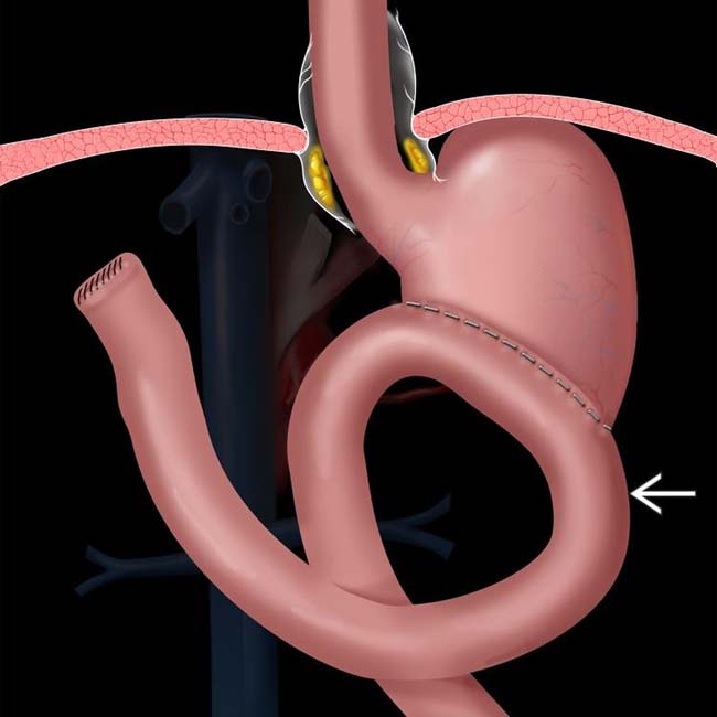
 enters the anastomosis from a left-to-right direction. This procedure is intended to reduce the prevalence of bile gastritis.
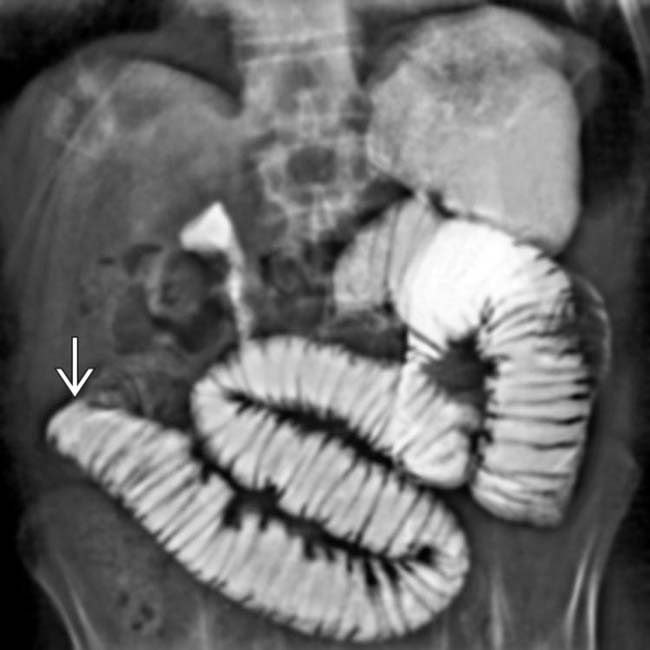
 . At surgery, a phytobezoar was removed, which corresponded to the shape and size of the gastric remnant.
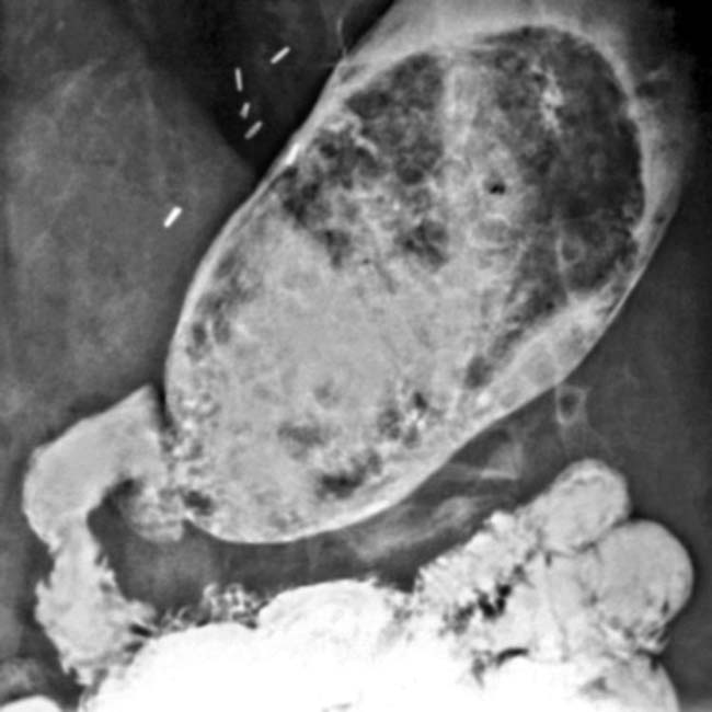


 Patients who have had partial gastrectomy for gastric cancer have high risk of recurrent tumor


