Partial Vertebral Duplication


Anteroposterior radiography depicts a typical supernumerary hemivertebra image 
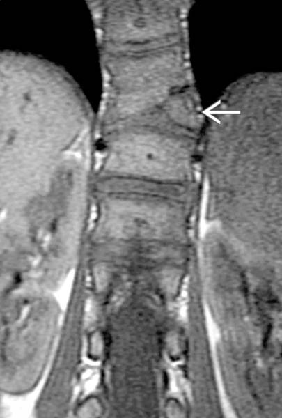
positioned between the right L2 and L3 vertebral levels, resulting in convex right scoliotic curvature.


image
Coronal T1WI MR depicts a small left thoracic supernumerary hemivertebra image 
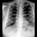
producing minimal convex left lower thoracic scoliosis.


TERMINOLOGY


Synonyms


• Supranumerary vertebra


Definitions


• Partial duplication of vertebral column → 1 or more supranumerary (“extra”) vertebra


IMAGING


General Features


• Best diagnostic clue
image Atypical scoliosis (sharply angulated, single curve, or focal) with 1 or more “extra” hemivertebra

• Location
image Thoracolumbar > cervical

• Size
image Hemivertebra is smaller than normal vertebra

• Morphology
image “Extra” hemivertebra frequently associated with focal scoliotic curvature


Radiographic Findings


• Radiography
image Supranumerary lateral hemivertebra with scoliosis


CT Findings


• Bone CT
image Supranumerary lateral hemivertebra


MR Findings


• T1WI
image Supranumerary lateral hemivertebra ± dysraphism, spinal cord anomalies

• T2WI
image Same as T1WI


Imaging Recommendations


• Best imaging tool
image Anteroposterior plain radiograph

• Protocol advice
image AP plain film for “counting” to localize abnormal vertebral level

image Long-cassette standing weight-bearing radiograph to quantitate scoliosis

image Multiplanar MR imaging to evaluate vertebral anatomy, assess for associated neurological abnormalities

image CT with 3D reconstruction: Excellent depiction of hemivertebra and scoliosis

Only gold members can continue reading. Log In or Register to continue

Stay updated, free articles. Join our Telegram channel

Oct 5, 2016 | Posted by in NEUROLOGICAL IMAGING | Comments Off on Partial Vertebral Duplication

Full access? Get Clinical Tree

Get Clinical Tree app for offline access