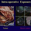
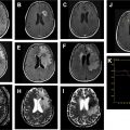
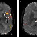
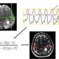
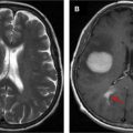
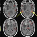
Functional magnetic resonance imaging (fMRI) is useful for localizing eloquent cortex in the brain prior to neurosurgery. Language and motor paradigms offer a wide range of tasks to test brain regions within the language and motor networks. With the help of fMRI, hemispheric language dominance can be determined. It also is possible to localize specific motor and sensory areas within the motor and sensory gyri. These findings are critical for presurgical planning. The most important factor in presurgical fMRI is patient performance. Patient interview and instruction time are crucial to ensure that patients understand and comply with the fMRI paradigm.
Key points
- •
Clinical functional magnetic resonance imaging (fMRI) is useful for localizing eloquent cortex in the brain prior to neurosurgery.
- •
With the help of fMRI, hemispheric language dominance can be determined, and Broca area, Wernicke area, and secondary language areas can be localized.
- •
The specific motor and sensory areas within the motor and sensory gyri also can be localized.
- •
The most important factor in presurgical fMRI is patient performance. It is crucial to be generous with patient interview and instruction time to ensure that patients understand and comply with the fMRI paradigm.
Introduction
Preoperative functional magnetic resonance imaging (fMRI) in patients with brain tumors or seizure disorder has moved into the clinical arena. , Clinical fMRI is very different from basic fMRI, however, and requires a specialized and dedicated approach. Unlike healthy volunteers for basic science research, patients in need of fMRI often present with significant impairments that complicate the examination. Developing patient-specific fMRI paradigms and extensively preparing patients for the scan are necessary to mitigate the effects of patient impairment. Thus, the most important aspect of fMRI administration occurs before a patient even gets in the scanner.
Scan preparation
Proper preparation is crucial to successful administration of clinical fMRI. When an ordering physician requests an fMRI examination, 4 pieces of information must be collected. These data should inform the approach taken in each case.
Age
There are documented differences in fMRI performance and blood oxygen–level dependent (BOLD) response between older and younger patients. Older patients may produce more head motion; patient age and motion have been positively correlated. Care must be taken to emphasize the importance of keeping the head still. Older patients show fewer activated voxels and greater noise in activated voxels, resulting in reduced signal-to-noise ratios. Several studies have shown that the BOLD response is lower in amplitude in older adults than in younger adults, which may reflect differences in neurovascular coupling. Working with older patients may require additional instruction or in-scanner guidance to mitigate contamination of the BOLD response with noise or motion artifact. Using aural and visual stimulation simultaneously during fMRI tasks may help these patients successfully complete the examination.
On the opposite end of the age spectrum, pediatric fMRI also requires additional preparation. The most common reasons for failure in pediatric scans include excessive head motion, insufficient instruction, and fear of the noise or confinement of the scanner. Younger children generally are less successful with fMRI. Children younger than age 8 years have shown significantly lower success rates than children over age 8 years. Even so, in-scanner head motion (relative mean displacement [mm]) may be 4 times higher in a patient aged 8 years compared with a patient aged 20 years. The effect of age on head motion seems to decrease with age. It is important to consider the particular challenges of scanning pediatric patients. Familiarizing children with the magnetic resonance imaging machine, providing ample instruction, and practicing before the scan may improve the quality of the results.
Handedness
The handedness of patients is important to consider when interpreting language fMRI cases. Many studies have shown that more than 90% of right-handed individuals (often as many as 95%) show left hemispheric (typical) dominance for language. Among left-handed individuals, however, as few as 74% show typical left dominance. The incidence of bilateral language activation also is higher among left-handed than among right-handed people. There may be a linear relationship between the degree of laterality and degree of handedness, with higher incidence of right hemispheric (atypical) dominance among the strongly left-handed than among the ambidextrous and strongly right-handed. ,
Because a relationship has been observed between handedness and language dominance, it is important to collect this information from the patient. Knowing a patient’s handedness informs interpretation of the fMRI results. For instance, if patients are left-handed, it is more likely that they show atypical language activation. The most common tool used to assess handedness is the Edinburgh Handedness Inventory. This measurement includes information about hand preference for everyday activities, including writing, drawing, and using scissors. Some older naturally left-handed patients may have been trained to predominately use the right hand. These forced right-handed individuals may show different functional neuroanatomy than naturally right-handed individuals.
Deficits
Because impairment can severely limit a patient’s ability to perform fMRI tasks, it is crucial to identify patient deficits prior to scan administration. Brain tumor patients may suffer from neurologic symptoms, including dysphasia, paresis, forgetfulness, confusion, and anxiety. Patients also may suffer from claustrophobia and may need premedication. Modifying existing paradigms is essential to accommodate many of these limitations. , A patient with hand or foot paresis likely will struggle to perform assigned motor tasks. In this case, the paradigm can be modified to apply external sensory stimulation to the affected area. This can be used to produce motor activation despite a patient’s motor impairment. , A patient with dysphasia or aphasia likely will have difficulty with standard language paradigms. Slowing the timing of the paradigm or reducing the frequency of stimulation can help improve patient performance. It also may be helpful to select the simplest task from the battery of language paradigms. Gathering information about functional impairment from either the patient or the ordering physician allows the fMRI to be tailored to each patient. Modifying the paradigm to suit each patient’s individual abilities gives the patient a greater chance of success.
Tumor Type and Location
When working with brain tumor patients, it is crucial to consider a patient’s cancer diagnosis and lesion location. High-grade gliomas, such as glioblastoma multiforme, may increase the risk of false-negative results. This is thought to occur through neurovascular uncoupling, in which the tumor disrupts normal neovasculature, such that an increase in neuronal activity does not lead to an increase in blood flow. , Because fMRI relies on normal BOLD response, disruption of the normal neovascular response can result in reduced fMRI activation. , , The radiologist interpreting the fMRI data in such cases must consider the risk of false negatives. The person administering the fMRI examination also must be aware of this risk to monitor the fMRI results effectively in real time.
Gliomas can alter the expected fMRI response due to cortical plasticity. Because low-grade gliomas grow more slowly than high-grade gliomas, these tumors may be more prone to functional reorganization. This slow rate of tumor expansion, as opposed to acute injury, may allow the brain to slowly reorganize and develop compensatory mechanisms. Intrahemispheric and interhemispheric reorganization both have been observed, , with activation seen in local preserved cortices and analogous contralateral structures, respectively. The possibility of functional reorganization may explain why most patients with low-grade gliomas show normal or nearly normal neurologic examinations. In such cases, when the left Broca area is invaded, for example, atypical right Broca activation may reflect compensatory activation. It, therefore, is important to consider how a patient’s diagnosis may affect the fMRI results, particularly when the results are unexpected.
Finally, the tumor location should inform paradigm selection. When a tumor is close to eloquent cortex, functional paradigms designed to activate that cortex should be performed (eg, language paradigms for tumors close to Broca area). Choosing appropriate paradigms based on different clinical scenarios is discussed later.
Patient evaluation
Patient Interview
Successful clinical fMRI requires detailed prescan patient preparation. This component of the examination requires interviewing, evaluating, and instructing the patient. , , , The interview often begins by assessing the patient’s clinical status, including asking about any deficits. The patient’s handedness also is determined. It is critical to evaluate the function that will be tested during the fMRI examination to ensure that the patient will be able to perform the task. As discussed previously, it is essential to modify any existing functional paradigms to accommodate patient limitations.
For a hand motor paradigm, for example, hand weakness must be tested for prior to the scan. For a language paradigm, the patient’s productive or receptive speech must be tested. The authors use the Boston Naming Test, a subtest of the Boston Diagnostic Aphasia Examination, to evaluate productive speech. The Boston Naming Test contains 60 standardized line drawings that the patient is asked to name, organized in order of word frequency. Auditory responsive naming questions describing concepts (eg, “name a big animal with a trunk”) may be used to test the patient’s receptive speech ability. Informal assessment through conversation also can provide evidence of possible deficit.
After the patient is evaluated, the procedure is explained to the patient in detail. This explanation should familiarize the patient with the task and the paradigm timing, including how the in-scanner instructions will be administered (ie, visually or aurally). For example, the patient should be told to expected to be still for 30 seconds and then tap fingers for 20 seconds, repeating this sequence 8 times. It is advisable to do a brief trial run of the selected task to maximize compliance in the scanner. , The patient’s success or lack thereof in the prescan rehearsal also can serve as a predictor of patient performance. If there are special circumstances, such as a visual impairment, hearing difficulty, or language barriers, these should be addressed during the prescan interview. , If a patient is receiving contrast, nursing staff should insert the intravenous line at this time.
The patient should arrive at least 1 hour before the scheduled scan to complete these tasks. This preparatory time spent with the patient can make the difference between a strong fMRI map and an uninterpretable one. Adequate patient preparation and evaluation are essential.
Language
fMRI paradigms should be selected based on a patient’s clinical scenario. These should include a thorough review of prior imaging and an evaluation of patient symptoms. Because the goal of presurgical fMRI is to identify eloquent cortex for the benefit of the neurosurgeon, the paradigms selected must seek to activate brain regions in close proximity to the lesion. This section focuses on paradigms designed to activate the language areas of the brain based on functional neuroanatomy. There are several situations in which it is appropriate to perform language fMRI paradigms.
It is important to identify hemispheric language dominance in cases where language areas are impacted. As discussed previously, most right-handed individuals display typical left hemisphere language dominance. In right-handed patients, language paradigms, therefore, are appropriate for most left hemisphere lesions; they also are appropriate for right hemisphere lesions when a patient displays aphasia. Because it is highly unlikely that a right-handed patient shows right hemisphere language dominance, it usually is unnecessary to perform language paradigms in these cases unless clinically indicated (aphasia). For left-handed and ambidextrous people, language paradigms are appropriate for lesions in both hemispheres.
Language Functional Neuroanatomy
The language network is composed of several essential primary language areas and additional secondary language areas. Although there may be variability among individuals in precise anatomic localization, , the frontal lobe generally controls productive speech and the temporoparietal lobes generally control receptive speech.
Primary Language Areas
Classically, the main productive speech area in the frontal lobe is known as Broca area. This area, located within the inferior frontal gyrus, and more specifically within the pars opercularis and pars triangularis (Brodmann areas 44 and 45), is essential for expressive language. Damage to this area can produce Broca aphasia, a nonfluent aphasia characterized by telegraphic, dysarthric, or agrammatic speech, or even mutism in severe cases. , , Patients may have difficulty with object naming, word fluency, and other productive components of speech ; however, comprehension commonly remains intact with insult to Broca area. Direct cortical stimulation (DCS) of this region during an awake craniotomy can produce total speech arrest. , Therefore, neurosurgeons commonly order fMRI examinations when there is tumor in the vicinity of Broca area.
Wernicke area, the main receptive speech area, is located in the posterior superior temporal gyrus posterior to the Heschl (auditory) gyrus. , This region classically has been considered the main area for language comprehension in the brain. As opposed to frontal lobe dysfunction, patients with temporal lobe dysfunction tend to present with varied language deficits. Injury to the Wernicke area can result in phonemic paraphasia (eg, substituting “pike” for “pipe”), semantic paraphasia (eg, substituting “train” for “car”), fluent aphasia (eg, word salad), circumlocution, or word-finding difficulty. , , Patients also may exhibit symptoms of receptive aphasia, including an inability to understand commands or answer questions. The varied nature of Wernicke aphasia aligns with DCS findings showing high variability of the localization of language function within the temporoparietal region. In 1 study in which DCS was performed on the temporal lobe, only 30.6% of patients showed a temporal lobe language site. Wernicke area may also play a role in perceptual, integrative, and verbal memory functions.
Broca and Wernicke areas are part of the classic model of language (the Wernicke–Geschwind model). Recent work suggests that the simple distinction between areas of speech production and speech comprehension may not reflect the true nature of language processing in the brain. Many researchers favor a more complex view. Yet identifying Broca and Wernicke areas still is useful in presurgical clinical fMRI. Modern models of language postulate that several language areas play a supportive role in the language network. These brain regions are called secondary language areas.
Secondary Language Areas
One well-known secondary language area is the middle frontal gyrus (MFG). , , , This region, also known as the dorsolateral prefrontal cortex, is located superior to Broca area. Language fMRI tasks can produce strong activation of the MFG that is comparable to Broca area in degree of lateralization. For example, strong left-sided Broca area activation may be accompanied by strong left-sided MFG activation, which supports left hemispheric dominance. The MFG is important for verbal working memory as well as cognition and information integration. DCS of the MFG infrequently produces language disturbance (approximately 10%) , ; however, dysarthria and anomia both have been observed with insult to the region. When resections have resulted in postoperative language deficits, these deficits generally have been temporary. ,
The supplementary motor area (SMA), despite its name, also plays a supportive role in language function. The SMA is located in the medial superior frontal gyrus, with the foot motor homunculus located just posteriorly. The SMA consists of 2 functional subdivisions, known as the pre-SMA (anterior-most portion of the SMA) and the SMA proper (posterior to the pre-SMA). , The pre-SMA and SMA proper are thought to support language and motor planning, respectively. There also is evidence of a central SMA, with joint motor and language function. , Resection of the SMA is known to produce SMA syndrome, a transient postoperative disorder characterized by a paucity of speech or akinetic mutism. Strangely, SMA syndrome often resolves on its own within weeks to months. , A patient is at 100% risk of developing SMA syndrome when a tumor is within 5 mm of the SMA. Resection of tumors in the SMA typically is considered safe due to the transient nature of the resulting postoperative deficits.
Another area of interest is the insula; however, this region’s role in language is not well understood. This is in part because it is a relatively deep structure, located deep to the frontal operculum. , A meta-analysis of fMRI studies has implicated the insula in several different components of language, showing similar insular activation for both receptive and expressive speech. This region likely plays a role in end-stage speech production (articulation) , and ventilation during speech. It also may play a part in naming, word-finding, and articulation. Damage to the insula may produce variable language deficits or none at all. Speech apraxia has been correlated with insult to the region. Damage to the insula alone is rare, which has made it difficult to draw conclusions about the region’s role in language. Although this region is not a target for presurgical fMRI, insular activation frequently is seen during language tasks.
The final language areas to consider are the supramarginal and angular gyri, which together comprise the inferior parietal lobule. The supramarginal gyrus is located anterior to the upturned aspect of the sylvian fissure, whereas the angular gyrus surrounds the posterior aspect of the sylvian fissure. Although the function of the angular gyrus is not well defined, a meta-analysis has shown that the angular gyrus frequently activates during semantic processing. The region may support reading, in particular. , On the other hand, the supramarginal gyrus may contribute to phonological processing and verbal working memory. There are limited surgical data for these 2 regions; however, the angular gyrus is related to alexia and agraphia, and the supramarginal gyrus is related to anomia and slurred/slowed speech. fMRI typically is not used to sensitively predict deficits based on the location of the inferior parietal lobule; rather, identifying reading-associated cortices often occurs via DCS during neurosurgery.
This discussion of primary and secondary language areas should serve as an introduction to the functional neuroanatomy of the language network. These areas can be expected to be seen to activate during language fMRI tasks. The greater understanding of the brain regions of interest, the more successful the presurgical fMRI.
Language paradigms
Paradigm Design
The fMRI paradigms selected should target the brain region of interest. The most common type of paradigm design is a block design, which consists of alternating active and resting blocks. For example, for a hand motor task, the patient alternates between 20 seconds of hand movement and 30 seconds of hand relaxation for several repeating cycles. Averaging the signal from at least 5 to 6 of these cycles increases statistical power and produces a stronger fMRI signal. On the other hand, event-related paradigms also consist of active and resting blocks but instead are designed to capture the neural response after a single stimulus event. These paradigms are composed of much shorter stimulus periods, usually less than 4 seconds long, with many more repetitions. Because block paradigms produce a strong average signal with greater statistical power, this design is preferred in patients for clinical fMRI. , ,
Frontal Paradigms
Language paradigms should target productive (frontal) and receptive (temporoparietal) speech areas. , It is important to perform multiple language paradigms per language case to confirm the accuracy of the results (preferably 2 to 3). A lesion in the frontal lobe requires frontal speech mapping. For these cases, tasks involving word generation, such as semantic and phonemic fluency, frequently are performed. Semantic fluency tasks present the patient with a letter and ask the patient to generate words that begin with the given letter. Phonemic fluency tasks present the patient with a category (eg, animals, colors, or vegetables) and ask the patient to generate words belonging to that category. , Another paradigm used to activate productive frontal areas is the verb generation task. This paradigm presents patients with a noun and asks patients to generate verbs associated with that noun. For example, for the noun “baby,” patients would generate words, such as “eats,” “sleeps,” “cries,” and “crawls.” The verb generation task produces results with greater specificity and less variability in hemispheric dominance than other tasks, perhaps because it requires more complex language processing. These productive speech tasks generally elicit activation of the receptive speech areas as well.
Temporoparietal Paradigms
Lesions in the temporoparietal lobes typically target the Wernicke area. This region often is more difficult to measure than the frontal region, even when using tasks specifically designed for receptive speech. , Language mapping for these lesions generally requires comprehension tasks, such as reading, listening, and sentence completion. , During the sentence completion task, the patient reads a sentence in which the final word has been replaced with a blank line. The patient must generate a word that logically completes the sentence. This task can be administered visually or aurally. When administered visually, it is possible to implement a button response with 4 possible answer choices. For example, “Bill gives haircuts and shampoos. He is a ________ (1) butcher (2) barber (3) batch (4) beer.” Another possible receptive task is auditory responsive naming. Patients listen to simple questions, such as “What do you shave with?” and “What color is grass?” and generate the answers to these questions.
Notes on Paradigms
Overlearned sequences, such as number counting, should be avoided in language mapping. Compared with object naming, number counting is a less sensitive task for both language localization and lateralization. Number counting has been shown to yield fewer areas of speech interruption during DCS than object naming, which may be due to the task’s simple, overlearned nature. Using one of the tasks, discussed previously, produces more reliable results. In certain cases, however, in which the patient has severe language deficits, number counting may be the only paradigm that the patient is able to perform. In the authors’ clinical practice, clinically meaningful fMRI language studies have been able to be acquired in such circumstances.
The fMRI patient typically is asked to generate responses silently (covert) rather than vocalize them (overt). Silent word generation during language tasks helps to minimize motion artifact due to vocalization; however, covert responses make it impossible to monitor patient performance aurally. Nevertheless, covert paradigms are suggested alongside real-time fMRI analysis to monitor patient performance.
Most fMRI paradigms elicit activation from both frontal and temporoparietal language areas. Despite attempts to isolate productive and receptive speech with specific tasks, many language paradigms activate Broca and Wernicke areas equally. , , It is possible to adapt most, if not all, language tasks to either aural or visual paradigm delivery. For some patients, one method works better than the other (eg, in cases of visual or hearing impairment). Aural delivery may allow for more dynamic administration of the examination, which is useful when adapting to patients’ limitations during the scan. On the other hand, visual delivery may produce more consistent results.
Lateralization
An essential goal of presurgical language mapping is to determine hemispheric language dominance. Intracarotid amobarbital testing (Wada testing) is considered the gold standard for determining language lateralization. During Wada testing, one of the brain’s hemispheres is anesthetized in order to test language function in the other. fMRI is a less invasive alternative to Wada testing because it does not involve sedation. Several studies have determined concordance between Wada and fMRI testing to be in the range of 86% to 91%. Discordance between the 2 methods may be caused by variability in fMRI acquisition, paradigms, and analysis, which can result in language maps of varying quality. Regardless, the high concordance and lower risk of fMRI have led many centers to embrace fMRI for presurgical language mapping.
Motor
As with language fMRI paradigms, motor fMRI paradigms must be selected based on a patient’s clinical scenario. This requires review of the patient’s symptoms and prior imaging to determine if the lesion impinges on eloquent motor cortex. Even if the motor cortex is not impacted directly, if a patient has motor or sensory deficits, it may be helpful to perform motor paradigms for the affected area (ie, hands, face, and so forth.). For some language cases, it also may be necessary to use motor paradigms to identify the tongue motor area.
Motor Functional Neuroanatomy
The primary motor cortex (M1) is essential for movement, and the primary somatosensory cortex (S1) is essential for sensation. Both motor and sensory systems are organized topographically; the motor and sensory functions of different body parts are located in specific locations along the precentral and postcentral gyri. These maps of motor and sensory function along the cortex are known as the motor homunculus and sensory homunculus, respectively. In the motor homunculus, the foot and leg motor areas are located along the interhemispheric fissure. The hand motor area is lateral to the foot motor area, and the tongue and face motor areas are lateral to the hand motor area.
The SMA is composed of the pre-SMA and the SMA proper, the latter of which is involved in motor planning and also is known to support word articulation. Because motor paradigms are necessary when a lesion impinges on the SMA, many patients undergo motor mapping to localize this region. This is important because the anatomic boundaries of the SMA are not well defined, which puts patients at risk for SMA syndrome.
The organization of the motor and sensory regions must be understood to properly plan for each fMRI, because tasks must be chosen based on the motor homunculus. More specifically, determining the location of the lesion within the motor strip determines which motor tasks should be performed. For example, a patient with a lesion in or near the tongue area of the primary motor cortex needs to perform a tongue motor task. Patients with lesions close to the midline should perform foot motor tasks, whereas patients with lesions close to the reverse omega portion of the central sulcus (the hand motor area) should perform hand motor tasks.
Ultimately, the purpose of fMRI motor mapping is to identify the motor areas that have a close anatomic relationship with the lesion. , Such motor mapping also is helpful when anatomy is ambiguous or effaced due to the presence of a lesion; the neuroradiologist may be able to identify the precentral and postcentral gyri in these cases with the help of fMRI.
Foot Motor
When the foot motor area is impacted, identifying the foot motor activation is critically important. Losing foot mobility can render a patient wheelchair-bound or bedbound. The foot motor task requires patients to wiggle the toes by performing toe flexion and extension. This task is somewhat difficult to perform without introducing movement, because patients often move the entire body in the z (inferior to superior) direction when wiggling the toes. Care must be taken to instruct the patient to isolate movement to the feet and toes. An alternative is to ask the patient to tap the feet against one another in the x and y directions (perpendicular to the long axis of the body). This also decreases motion in the z direction.
Hand Motor
The hand motor (finger-tapping) task requires patients to tap their thumbs to each finger sequentially while minimizing other body movements. If patients suffer from distal hand weakness, they can perform a modified version of the task in which patients simply open and close the hands (fist clenching). , If a patient is too weak to complete the modified motor task, passive hand simulation should be performed. When a patient has weakness in 1 hand, the activation map may be asymmetric. It often is useful to perform the motor task first with both hands and then with the affected hand alone in order to focus on the affected hemisphere.
Tongue and Face Motor
To elicit tongue motor activation, patients are asked to keep the mouth closed and to sweep the tongue against the back of the teeth, taking care to avoid head motion. Only a slight amount of movement is necessary to activate the tongue motor area because the tongue, mouth, and lips take up a relatively large portion of the motor homunculus. If lip or face motor activation is of interest, patients are asked to purse the lips and push them back into a smile, repeatedly. Locating the tongue and lip motor areas often takes place with language mapping.
Passive Stimulation
As discussed previously, when a patient is too weak to perform the hand or foot motor task, passive sensory stimulation must be applied in the form of brushing, squeezing, or touching the hand or foot. It has been shown that passive stimulation is a valid alternative or complement to active motor paradigms. Passive stimulation also is useful when a patient is unable to follow the paradigm instructions or complete the motor task without excessive head motion. Alternatively, if the postcentral gyrus is impacted or if a patient suffers from numbness, performing sensory paradigms may be useful. Although sensory paradigms activate the postcentral (sensory) gyrus, they often activate both motor and sensory areas. , Because such reciprocity exists between motor and sensory areas, the results of these tasks must be interpreted with caution.
Scan set-up
After assessing the patient’s clinical situation and completing the prescan interview, the final step before administering the fMRI is setting the patient up in the scanner.
Which type of paradigm delivery system to use during the scan must be decided. For visual delivery, a back projection and mirror may be used. A sophisticated set of noise-canceling headphones and liquid crystal display goggles for visual delivery also can be purchased. For aural delivery, instructions can be delivered to patients through headphones or in-room speakers. As a last resort, if other methods fail, the magnetic resonance (MR) room can be entered and the patient’s leg or arm simply tapped to indicate “start” and “stop.” It is essential to test the system prior to beginning the scan. The patient must be able to hear what is being said through the headphones and see what is being presented visually on the screen or goggles.
It is important to remind the patient that the scan is extremely sensitive to motion and that head motion must be minimized. Ensuring that the patient is in a comfortable position in the scanner increases compliance and reduce motion. , The patient can be made more comfortable by putting a pillow under the knees and providing a blanket. Patients also should use the restroom before the scan begins so that they are able to complete the entire study without pause. This is true especially for children. Patients must stay in the same position for the entire study, even when the scanner is not acquiring images, so that the fMRI images can be coregistered onto the high-resolution postcontrast T1-weighted images. To make this possible, patients must minimize all motion.
Once a scan begins, it is useful to speak to the patient through the headphones between sequences. A small amount of encouragement can go a long way. Because the fMRI paradigm is not intuitive to most patients, reiterating instructions immediately before each task can increase compliance. In addition, reassurance that they are performing the task correctly may improve patients’ performance. Between each sequence, the task also can be clarified or adjusted as needed.
Monitoring patient performance is an important part of administering the fMRI examination. For example, if a patient is asked to perform finger-tapping, the patient must be watched to ensure that the patient actually moves the fingers. If the patient fails to perform the task, the results are meaningless. Most MR machines allow the operator to monitor fMRI activity in real time. This is a useful tool for monitoring patient performance, especially for covert language tasks, which are restricted to the patient’s mind. If the language regions are visible on this raw map, there can be fair confidence in the quality of the fMRI results.
Summary
Clinical fMRI is a useful tool for localizing eloquent cortex in the brain prior to neurosurgery. Language and motor paradigms offer a wide range of tasks to test specific brain regions in the language and motor networks. With the help of fMRI, hemispheric language dominance can be determined and Broca area, Wernicke area, and secondary language areas can be localized. It also is possible to localize specific motor and sensory areas in the motor and sensory gyri. These findings are critical for presurgical planning. The most important factor in presurgical fMRI is patient performance. Sophisticated fMRI devices and software offer little benefit if time is not taken to prepare for the scan, examine the patient, explain the procedure, make any necessary adjustments, and monitor the patient throughout the examination. It is crucial to be generous with the interview and instruction time to ensure that the patient understands and complies with the fMRI paradigms. With proper patient instruction and paradigm selection, the battle for quality fMRI results already is half-won.
Clinics care points
- •
Pre-operative brain tumor patients often neurologically compromised and may have difficulty understanding or performing the fMRI paradigm.
- •
A neurological examination and patient preparing the before the fMRI scan is essential.
- •
The most important determinate of a successful fMRI examination is patient cooperation.
- •
Monitoring the patient during the fMRI examination to ensure compliance with the paradigm will increase the number of fMRI studies of diagnostic quality.
Acknowledgments
Funding support provided by the National Institutes of Health (NIH) : NIH-NIBIB R01 EB022720 (Makse and Holodny, PI’s); NIH-NCI R21 CA220144 (Holodny and Peck, PI’s); NIH-NCI U54 CA137788 (Ahles, PI); and NIH-NCI P30 CA008748 (Thompson, PI).
Disclosure
N.P. Brennan and M. Gene have nothing to disclose. A. Holodny is the Owner/President of fMRI Consultants, LLC, a purely educational entity.
References
Stay updated, free articles. Join our Telegram channel

Full access? Get Clinical Tree


