Its own capabilities and limitations
Capabilities (and limitations) of alternative procedures
Clinical questions:
Diagnosis, prognosis, connection with therapy
Local scenario:
Instruments and procedures routinely used
Epidemiological and socio-economic issues
Risks and prejudices
1.3.1 General Capabilities of NM
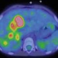
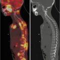
In the paragraphs above, we described how effective nuclear medicine can be because of its capability to produce a molecular and/or pathophysiological imaging. Furthermore, with respect to other procedures, as the US, NM is advantaged because it is reproducible and not operator dependent. This point in favour is also accompanied by a panoramic view, not allowed by US. Furthermore, mainly in case of positive indicators, as FDG and Tc-99 m diphosphonate, showing a more intense uptake in pathological tissues with respect to the normal ones, a whole-body scan, very helpful in staging and restaging, may be acquired. Using radionuclide techniques is also standardizable and therefore more reliable quantitative analyses, allowing a better evaluation of nonfocal diseases and/or a more precise definition of changes which appear in the follow-up, eventually as response to therapy.
A further positive issue connected with NM is dependent on the capability to define a prognostic information, as it happens with FDG in oncology. Using radiocompounds, as FDG or radiolabelled white blood cells (WBC), is also possible to define disease activity in many chronic inflammatory conditions, finding a relevant role in recruiting only patients that may successfully undergo to therapies. With respect to therapeutic strategies, a major advantage may be acquired in the presence of radiopharmaceuticals that may be labelled either with γ or β + emitters and with β-radionuclides, as it happens for radioiodine, metaiodobenzylguanidine (MIBG) and somatostatin analogues. Using this approach, it is possible to forecast a therapeutic efficacy, on the basis of an a priori evaluation obtained with a similar radiocompound, administered at a significantly lower radiation dose. As reported above, the complementary contribution given in prognosis and therapy may create a clinical indication for NM also as second-line diagnostic procedure after a first “pathological” diagnosis has been already obtained.
1.3.2 General Limitations of Nuclear Medicine
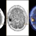
Being based on difference of concentration and not of density, NM cannot give an anatomical information. Moreover, radionuclide procedures don’t allow a loco-regional staging, mandatory before a surgical choice, as an example to individuate relationships between mass and adjacent vessels. Many of these limitations have been recently solved by the availability of hybrid machines, including PET/CT, SPECT/CT and more recently PET/MRI, which permitted a significant increase in accuracy, either decreasing false-negative or false-positive results, with respect to the individual procedures considered alone.
A major limitation associated with NM is certainly related to the presence of ionizing radiations, a disadvantage shared with CT and traditional Rx. Nevertheless, in the presence of an effective clinical indication, there are no absolute contraindications for radionuclide techniques, although radioisotopic procedures have to be always “justified”. It means that no scintigraphies or PET or SPECT studies may be performed when alternative procedures permit the achievement of a similar information without radiations. This rule is more restrictive in paediatrics (and even more in pregnant women), being the stochastic risk associated with nuclear medicine conditioned either by the percentage of cells that multiply, higher in infancy, or by the life expectancy, longer for paediatric patients. It has however to be pointed out, as we will see below in the paragraph evaluating risks, that the calculation of a cost/effective balance is not always easy, mainly in comparison with MRI, negatively affected by a minor diffusion and frequently accompanied by higher costs and by a high rate of studies non-executable in paediatrics without narcosis.
As a negative counterpart for nuclear medicine, it has to be remembered that problems for radioprotection may be increased considering the radiation charge for physicians, nurses, technicians and relatives or other caregivers, the presence of which may be requested to facilitate the procedure. In this sense, although the dose of radiation and an increasing incidence of cancer are typically very low, a justification is mandatory both for the patient and for accompanying persons.
1.4 Technical Problems of NM in Paediatrics
Although they are not exclusive of paediatric patients and not present in all the subjects, also because of the wide differences existing, for example, in early childhood with respect to the adolescence, some technical problems are peculiar in this population; they may be due to factors such as the body’s structure and size; difficulties in injecting radiopharmaceuticals, due to the small calibre and fragility of the vessels; inability to collaborate which may cause disturbing movements or an increased risk of contamination; psychological structure frequently governed by fear of the unknown; and so on.
While it is impossible to exclude ionizing radiations from radionuclide studies, to perform a study allowing an effective clinical response at the lowest cost, which has also to consider risks and the reliable solution of technical problems, it has to be a professional duty [3].
1.4.1 How to Approach the Paediatric Patient in Nuclear Medicine
In paediatric imaging, a successful diagnostic examination is obtained when the achievement of quality images, without degradation due to technical problems, occurs without mental or physical detriment to the patient. The ability of a child to remain sufficiently immobile during the scan depends upon his or her behaviour and the administered technique itself. Infants and small children are unable to cooperate and to follow verbal directions. Many older children are cooperative with adequate support and guidance during the exam.
It has to be remembered that paediatric patients in the NM department are often subjected to unexpected procedures that cause pain and increased anxiety and distress, like intravenous or subcutaneous injections and urethral or angiocatheter insertions. The use of topical creams to provide topical anaesthesia has been shown to reduce the pain associated with these procedures. Conversely, anaesthesia has to be avoided as much as possible, because, although it may allow a “technically perfect” scan, it is dangerous and expensive. Furthermore, it may negatively affect the examination conditioning the pharmacokinetic of the injected radiotracer. Similarly, sedation has to be performed only exceptionally and when absolutely needed, because of serious associated risks, such as hypoventilation, apnoea, airway obstruction, laryngospasm and cardiopulmonary impairment. These adverse reactions, which may occasionally occur during and/or after sedation, can be minimized with a procedure carefully performed, but not completely eliminated.
Children’s weight varies from premature neonates, weighing less than 1 kg up to 100 kg and more in teenagers. This condition creates a huge diversity in physiology, pathology and psychology. Therefore, starting from the arrival of the patient in the department, a sufficient time is needed to allow an individual assessment, based on many issues as an interactive discussion in acquiring a consent, including the activation of special preparation procedures to the exam, such as a play therapy.
When possible, information about the procedure should be given beforehand through information sheets sent to the family or through a phone call with preparation instructions. In general parents, or other close relatives such as grandparents and uncles, may better help the children when they are prepared as well. Therefore the procedure has to be explained to the parent (and/or to the alternative caregiver), and any question or concern has to be addressed as required. It is essential to give to the accompanying person the sufficient time to ask questions or express concerns at any point, particularly when one is dealing with frightened or anxious children, who may be less cooperative if they do not understand what is happening to them. Conversely, it is important to restrict the number of interacting relatives, individuating only one or two of them as possible caregiver, to avoid confusion and the activation of negative behaviours [4].
A child-friendly approach and patient preparation are major issues for the success in the large majority of nuclear medicine procedures. Children should be prepared for what they will face, to lessen their anxiety and promote their cooperation. Such preparation should be based upon the developmental level of the child. The role of the parent should be supported when possible. Most kin and children have a desire to be together during procedures. Policies should be developed to offer this opportunity. The presence of a parent is comforting to a child and can lessen anxiety. Allowing a protective person to remain in the room during the scan time can also give the child a sense of security, helping an otherwise uncooperative subject to successfully complete the scan without the need for sedation. It can be also helpful to allow the little patient to bring a favourite toy or stuffed animal into the scanning room, if possible. This toy can be placed above the head of the subject or held in his or her hands, out of the field of view.
Children need to know what will be required to them to gain their cooperation. Therefore they should be prepared for the experience they will encounter in the nuclear medicine department. They should be given an age-appropriate explanation of what they will feel, hear, see and/or taste. Medical and paramedical personnel should provide encouragement and ample praise. The subjects should be approached with the positive expectation of success, to increase the rate of cooperative scans.
Distraction is a commonly used non-pharmacologic pain and fear management technique used by both healthcare professionals and parents to attenuate procedural hurt and distress. Distraction operates on the assumption that, by shifting a child’s focus to something engaging and attractive, his or her capacity to attend to painful stimuli is hindered. Thereby pain, distress and anxiety are reduced. A number of behavioural distraction techniques, such as watching a movie, listening to a story, or listening to music, can increase the child’s ability to tolerate the examination. Natural sleep in infants can be induced by food, comfort and warmth and represents a condition greatly facilitating the scan.
When restraining a child, it is important not to use excessive strength; the used force should be appropriate to the child’s age. The safety of the staff restraining a strong patient is also paramount to good practice. Training of professionals in effective risk minimization when restraining should be given. As with any paediatric procedure, intravenous access can be problematic depending on patient cooperation and hydration status. Establishing an intravenous line before injection allows the little patient time to recover, as the experience can often be painful and stressful. All the personnel involved with the patient should be familiar with the patient’s positioning, having also knowledge on the scan’s duration. Medical equipment and patient intravenous lines have to travel safely with the patient through the scanner, to maintain the patient safety and to have the capability to intervene, if necessary.
Once scanning is complete, images should be reviewed before the patient is transferred off the scanning bed, to ensure that no further imaging is required.
The management of uncooperative children should take into account their individual needs and fears, within the context of the illness, and in partnership with the parents or guardians. Ideally, the wishes of the child should be respected, and, if a competent subject is resistant to the persuasive powers of parents and professionals, the investigation must be delayed and reassessed [5].
1.4.2 The Paediatric Environment in Nuclear Medicine
If the disease “scares”, this happens even more frequently for younger patients, who have a greater fear of the unknown. In this sense, it is very important to create a familiar environment, where colours, lights, waiting rooms and tools of distraction, including televisions, toys, cartoons and so on may play an important role in creating an atmosphere of relaxation, in which the smiling staff professionalism is a fundamental added value. Of course, an important element favouring this goal is determined, as widely explained above, by the communication with the patient, when big enough to understand, and/or with his or her relatives. If part of the fear is connected with the unknown, the a priori knowledge of the steps that will be lived in the next few minutes or hours can certainly increase the collaboration of the young patient. As previously reported, it is very important to have interactive connection with the relatives that have to be tranquilized and eventually may be authorized to keep company to the kid after correct information of risks associated with ionizing radiations. In this sense, while a pregnant mother should never get in the authorized “hot” area, the cooperative participation of grandparents has to be stimulated with respect to the presence of younger caregivers. Of course, the contribution of nurses, technicians, physicians and/or other professionals involved may be requested, if needed. Considering that fear, pain, family dynamics, previous experience with diagnostic and therapeutic strategies can determine problems, it’s important to work for the best understanding of the procedure, trying to determine the more strict cooperation between all the actors of the study, first of all with the little patient. A psychological expertise by the physicians and professionals involved is very important, because information of the patient and of caregivers may also become detrimental, mainly in case of anxious subjects. A mandatory rule is never leave the children unattended.
1.4.3 Patient Preparation
With respect to the intravenous injection, the most important rule is never inject radioactive if you’re not sure you’re in the vein. To reach this goal, strategies utilizing butterfly needles and/or three-way catheters are helpful, and these operations have to be performed in the more relaxed situation, before the injection of the radiopharmaceutical. Of course, this suggestion is particularly critical when dynamic studies have to be acquired. We will not discuss here, devoting this information to following specific chapters, other invasive and painful procedures, such as the urethral catheterization in radionuclide cystography. We want only to remember that all the strategies having as their aim the reduction of pain and/or of risks of infection have always to be adopted. In this context, have also to be evaluated conditions that may reduce radiation dose to the patient, as those related to hydration and urinating. The risk of contamination has to be avoided using impermeable sheets. Similarly, the need of fasting; the knowledge of haematochemical data, as glycaemia before a PET-FDG scan; and the eventual relevance of the suspension of a therapy have to be well known before the radiocompound’s administration.
Related posts:
Stay updated, free articles. Join our Telegram channel

Full access? Get Clinical Tree


