1. Apnea
2. Full-term infant less than 1 month of age (unless an inpatient admitted to the hospital)
3. Respiratory compromised patients
4. Uncontrolled/unpredictable gastroesophageal reflux or vomiting that poses an aspiration risk
5. Craniofacial abnormality that may make it difficult to establish effective mask airway
6. Cyanotic cardiac disease or unstable cardiac status
7. Painful procedure that may be challenging to provide adequate analgesia without a general anesthetic
8. High-risk procedure that may require presence of an anesthesiologist for resuscitation
9. Procedure that requires absolute immobility only achievable with a general anesthetic
10. Procedure being performed in remote location that is so removed that immediate emergency backup assistance would be virtually impossible
11. Inadequate qualified personnel available to provide safe procedural sedation
The patients scheduled for non-anesthesio-logist-delivered sedation are typically American Society of Anesthesiologists level 1 or level 2 but rarely level 3 (Table 3.2).
Table 3.2
ASA physical status classification
1. A normal healthy patient |
2. A patient with mild systemic disease |
3. A patient with severe systemic disease |
4. A patient with severe systemic disease that is a constant threat to life |
5. A moribund patient who is not expected to survive without the operation |
6. A declared brain-dead patient whose organs are being removed for donor purposes |
In the United States, the delivery of deep sedation by non-anesthesiologists should be administered according to the American Society of Anesthesiologists Guidelines [3]. Under these guidelines, registered nurses are no longer qualified to administer deep sedation. The sedation provider must be a physician, nurse anesthetist, or anesthesia assistant. Specifically, the sedation care provider is expected to be experienced in the delivery of positive pressure ventilation via a facemask, endotracheal intubation, and insertion of laryngeal mask airways along with placement of nasal and oropharyngeal airways. A minimum of 35 patients or simulated cases must be performed in order to demonstrate competence. Recently, the Center for Medicaid and Medicare Services published their guidelines which are consistent with the American Society of Anesthesiologist’s qualification requirements for sedation providers [4].
In the event that there are questions regarding the medical status or appropriateness of a child for sedation, a liaison from the Department of Anesthesia can facilitate the evaluation process by reviewing the history and, if appropriate, requesting consultations with appropriate specialty services (cardiac anesthesia, otolaryngology, surgery, nephrology, cardiology, or endocrinology). If the patient is deemed appropriate to receive sedation, then personal discussion between the consulting physician and sedation care provider is helpful to maximize safe care.
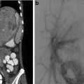
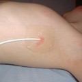
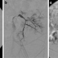
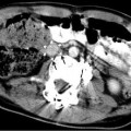
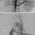
It is important for the practitioner responsible for sedation to have a thorough understanding of the procedure requested. For example, the same patient may be medically appropriate to undergo sedation for an MRI scan but may be an inappropriate sedation candidate for a nephrostomy tube, placed with the patient prone in interventional radiology. Although a patient may meet the medical criteria for sedation, a collaborative discussion between the practitioners responsible for the sedation and the radiologist will ensure that the procedure and patient lend itself to sedation. In the event that the procedure is deemed high risk (e.g., cerebral embolization), associated with significant pain (e.g., sclerotherapy with doxycycline), or long in duration, the patient is usually best referred to general anesthesia for management. Additional, important, considerations should be the physical layout of the procedure room and its geographical proximity to the operating rooms. In the event of an emergency and the need for a “code team,” the physical layout is important. If the radiological suites are physically isolated and distant from backup assistance, the more conservative approach will be to request anesthesia services prior to the initiation of a high-risk procedure.
Patient Sedation Guidelines
To minimize the chance of drug delivery error or miscalculation, it is helpful to have preprinted order sheets that should be approved by the Hospital Sedation Committee as recommended by JCAHO. The practice standards adopted by the American Society of Anesthesiologists in 1986 for basic intraoperative monitoring apply as well to extramural locations. Practice standards and guidelines promulgated by the American Academy of Pediatrics [5] are exceeded by established practice standards in anesthesiology [6]. Significant variances may exist when non-anesthesiologists sedate [7]. Practice Standards for Nonanesthetizing Locations were adopted by the American Society of Anesthesiologists in 1994 [8]. Recent practice standards by the American Society of Anesthesiologists for deep sedation by non-anesthesiologists stipulate monitoring which includes electrocardiogram, pulse oximetry, capnography, and noninvasive blood pressure monitoring [3].
A director of anesthesia services for a busy extramural radiology site will facilitate the delivery and coordination of anesthesia and sedation services. By being available to answer questions, do on-site consults, examine patients, and provide backup support or emergency airway expertise, the anesthesiologist is critical to the viability of a non-anesthesiologist-delivered sedation program. In addition to the American Society of Anesthesiologist, the Joint Commission on Accreditation of Healthcare Organizations (JCAHO) Anesthesia and Sedation Manual has set guidelines for credentialing of all personnel who administer sedation [3, 9].
Medications
The selection of a sedation agent depends on the patient’s underlying medical condition, age, drug tolerance, and anticipated procedure. Each medication has its own property which can include a hypnotic, anxiolytic, and/or analgesic. In order to appropriately make a sedation plan for each procedure, it is important to understand the properties of each medication and its potential synergistic actions with adjuvant medication. The more common sedatives administered for light, moderate, and deep sedation will be reviewed below.
Ketoralac
Ketoroloac (Toradol; Abbott labs, N. Chicago, IL) is an analgesic. It does not have sedative, hypnotic, or amnestic properties. Ketorolac tromethamine can be administered intravenously every 6 h with a maximum of 72 h of administration. It is useful for onetime administration to provide analgesia for short procedures such as biopsies. As a nonsteroidal anti-inflammatory agent, ketorolac may inhibit platelet aggregation and prolong bleeding time, which may be an undesirable effect for some interventional procedures. Agreement to administer a nonsteroidal should be obtained from the interventional radiologist prior to administration. Alternative analgesics could include narcotics or ketamine.
Narcotics
The choice of narcotic should depend on the duration of the procedure and the extent of analgesia required. Morphine (Baxter Healthcare Corp, Deerfield, IL) and fentanyl (Baxter) are the more popular narcotics. Morphine requires approximately 10 min for effect and can provide analgesia for up to 2 h. Fentanyl works within minutes and has 100 times the potency of morphine. It generally needs to be redosed every 30–60 min, depending on the procedure. Narcotics work best when administered prior to (in anticipation of) the painful stimulus so that adequate analgesia is present at the time of the stimulus.
Ketamine
Ketamine, 2-(o-chlorophenyl)-2-(methylamino) cyclohexane, a phencyclidine and cyclohexamine derivative, was developed and introduced into clinical anesthesia practice in the 1960s. It may be administered via intravenous, intramuscular, oral, rectal, nasal, epidural, or intrathecal routes. The use of ketamine for pediatric sedation and analgesia has been described in various nonoperating room settings that include emergency departments [10], gastroenterology [11], oncology [12], dental [13], and radiology suites [14, 15]. Ketamine produces rapid onset of deep sedation and analgesia with minimal respiratory depression and cardiovascular side effects [16–21]. A review of the literature reveals that despite the widespread use of ketamine by non-anesthesiologists, there is no consistent protocol for ketamine administration. Mason et al. describe the intravenous or intramuscular administration of ketamine, up to 2 mg/kg, for interventional radiological procedures. The intravenous dosage is followed by a continuous infusion of up to 125 μg/kg/h ketamine until the procedure is completed [17, 18]. When given in small bolus doses, it provides analgesia for an average of 30 min. As an infusion, ketamine can produce a continuous state of analgesia that may be titrated up and down in response to (or in anticipation of) the painful stimulus. It is especially useful for patients who are going to undergo an exceptionally painful procedure (doxycycline sclerotherapy, chest tube), are on chronic opioids, or have a high tolerance to opiates. The coadministration of ketamine with an anticholinergic is no longer recommended [19]. Ketamine provides an effective alternative to narcotics in some patients.
Hallucinations, delusions, nightmares, and emergence delirium are phenomenon most commonly described as a potential side effect of ketamine; these are more commonly noted in adults [22, 23]. The presence of these adverse events in the pediatric population is controversial [13, 24]. In adults, the concomitant administration of benzodiazepines (midazolam or diazepam) with ketamine has been shown to decrease the incidence of these events. Again, the utility of benzodiazepines in reducing these events in children is controversial [25–27]. Some reports indicate that the addition of benzodiazepines leads to an increased incidence of oxygen desaturation events [28]. Under age five, there is no definitive evidence that benzodiazepine administration will reduce the hallucinations, delusions, and excitatory behavior that can occur with ketamine. Children over age five may in fact benefit from concomitant benzodiazepine administration. Although ketamine administration and supervision by interventional radiologists is not a widely recognized technique, it offers an alternative agent for children who require profound analgesia albeit the risk of dissociative side effects.
Collaboration between the Departments of Anesthesiology and Radiology can provide for the radiologist-supervised administration of ketamine by nurses for interventional procedures, liver and renal biopsies included, in pediatric patients who might otherwise have required a general anesthetic. With sufficient triage and careful review of the medical history as well as relative and absolute contraindications to ketamine administration, radiologists can safely incorporate ketamine into their sedation armamentarium (Tables 3.3 and 3.4). Patients and parents are often relieved and grateful to avoid general anesthesia.
Table 3.3
Exclusion criteria for ketamine induced sedation
1. Active pulmonary infection or disease |
2. Known or potential (i.e., risk of) airway compromise |
3. Pulmonary hypertension |
4. Age of 3 months or younger |
5. History of apnea or obstructive sleep apnea |
6. Craniofacial defect that would make mask ventilation difficult |
7. Complex cardiac disease |
8. Acute globe injury |
9. Prior adverse reaction to ketamine |
10. History of bipolar disease or schizophrenia |
11. Head injury associated with loss of consciousness, altered mental status, or emesis |
12. Intracranial hypertension (i.e., CNS mass lesions, hydrocephalus, head injuries associated with increased intracranial pressure): if there is any doubt, please have radiologist consult ordering physician to determine whether there is increased intracranial pressure risk |
13. Any child in whom there is a question of increased intracranial pressure |
14. Child with potential ventriculoperitoneal shunt malfunction |
15. Increased intraocular pressure |
16. Patient or parent refusal |
Table 3.4
Adverse events and parent and patient satisfaction
Variable | Total cohort (n = 65) | Liver biopsy (n = 35) | Renal biopsy (n = 30) |
|---|---|---|---|
Adverse events (no. of patients) | |||
During sedation | 2a | 0 | 2 |
During recovery | 8b | 6 | 2 |
24 h after procedure | 4c | 2 | 2 |
7 day after procedure | 0 | 0 | 0 |
Sedation failure (no.) | 0 | 0 | 0 |
Satisfaction (%)d | |||
Dissatisfied | 0 | 0 | 0 |
Neutral | 8 | 6 | 10 |
Satisfied | 58 | 60 | 57 |
Very satisfied | 34 | 34 | 33 |
Preparing for Emergencies in Areas Distant to the Operating Room
There are three particularly challenging scenarios that may occur in nonoperating room (off-site) locations: (1) the child with a known difficult airway, (2) the child with an unrecognized difficult airway, and (3) cardiovascular arrest. Each challenging situation will be addressed in order and in detail below. Most interventional radiology suites are remote to the operating room, to skilled and knowledgeable airway (otolaryngology, anesthesia, and surgery) assistance, and to backup airway support (bronchoscopes and alternative airway devices). Fiber-optic equipment and procedures are not routine in outfield areas and thus, when anticipated, should be reserved for the operating room environment. It is suggested that the “potential difficult airway” be managed first in the operating room and, after the airway is secured, the patient can then be transported to the radiological suites.
In the operating room, there are appropriate backup support and supplies if needed. Remember that each extramural anesthetizing location is unique with regard to conducting resuscitation. Redundancy of monitoring devices and equipment is important; one should not be limited to a single item that could malfunction at the time of resuscitation. The physicians, nurses, anesthesiologists, technologists, and support personnel must know the location of emergency equipment. In addition, a hard board to be placed under the patient during resuscitation should be readily available. Mock codes and simulation of emergencies should be performed regularly to ensure adequate flow, teamwork, and delineation of responsibilities.
The administration of iodine-containing contrast for interventional procedures necessitates a complete knowledge of risk factors for anaphylaxis and appropriate prophylaxis and intervention (Table 3.5). Patients with multiple allergies, shellfish allergies, or atopic disease are at increased risk of exhibiting anaphylaxis to iodine-containing contrast. These patients may benefit from pretreatment with steroids and antihistamines.
Table 3.5
Management of acute reactions in children
Urticaria |
• No treatment needed in most cases |
• Give H1-receptor blocker: diphenhydramine PO, IM, or IV 1–2 mg/kg, up to 50 mg |
• If severe or widely disseminated, give ∝-agonist, epinephrine SC (1:1,000) 0.01 mL/kg |
