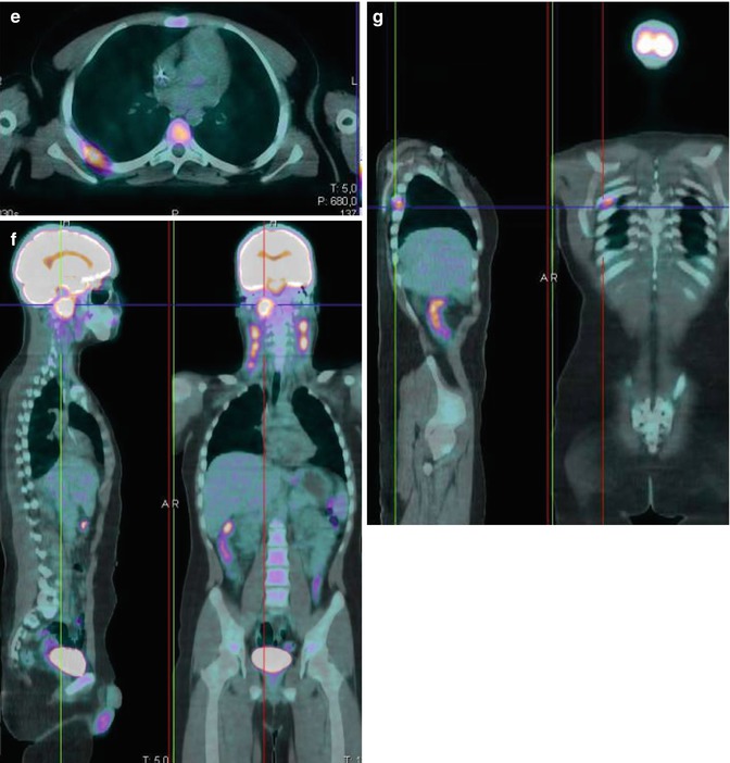
Fig. 13.1
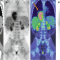
Baseline 18F-FDG–PET/CT scan shows intense uptake in the nasopharyngeal primary lesion (a, b, f) as well as in multiple bilateral enlarged cervical lymph nodes (c, d) and in a metastatic right pleural lesion (e, g)
The patient then underwent two cycles of chemotherapy (cisplatin and fluorouracil). His early response to chemotherapy was evaluated with ce-CT and 18F-FDG–PET/CT. Both examinations highlighted the positive response to chemotherapy. However, whereas several cervical lymph nodes >10 mm (arrow in Fig. 13.2) and persistent posterior right pleural thickening were still evident on the ce-CT, 18F-FDG–PET/CT showed a complete metabolic response (Fig. 13.2).
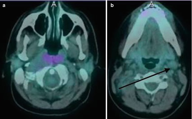
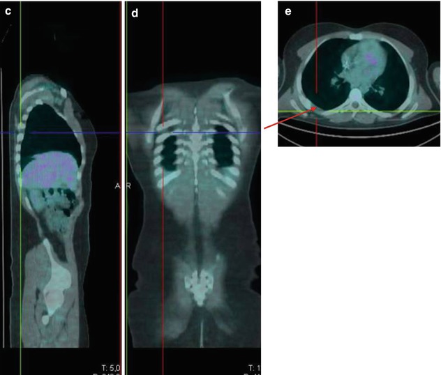


Fig. 13.2


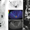
18F-FDG–PET/CT performed after two cycles of chemotherapy shows a complete metabolic response of the all pathological uptakes (a–e). On the opposite, enlarged cervical lymph nodes (black arrow in b) and posterior right pleural thickening were still evident on the coregistered CT images (red arrow in e)
Stay updated, free articles. Join our Telegram channel

Full access? Get Clinical Tree


