Pediatric PET/CT

IV
Nononcologic Applications


26
Pediatric PET/CT

M. Beth McCarville
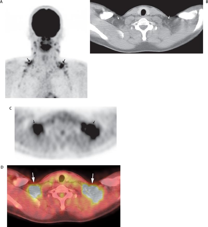

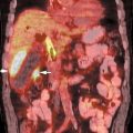
The use of positron emission tomography/computed tomography (PET/CT) in children requires consideration of several unique technical and logistic issues. Young children often require sedation for PET/CT, and personnel trained in the management of children are vital to patient safety. Pregnant guardians of young patients should not be allowed in the fluorodeoxyglucose (FDG) uptake room, and arrangements must be made for the supervision of such patients. Before administration of the radioisotope, young female patients themselves must be questioned regarding their childbearing potential. This delicate subject is best handled by a technologist with experience working with young girls. Additionally, interpretation of pediatric PET/CT imaging requires familiarity with pediatric anatomy and physiology because, compared with adults, children have more metabolically active brown fat and less retroperitoneal fat (Fig. 26.1).1 PET/CT is becoming an increasingly important adjunct to the care of the pediatric oncology patient. Several indications for its use in children are discussed herein.


image

Fig. 26.1 A 16-year-old boy previously treated for Hodgkin lymphoma. (A) Maximum intensity projection positron emission tomography (MIP PET) image showing intense fluorodeoxyglucose (FDG) activity in the supraclavicular areas (arrows) that is difficult to accurately localize by positron emission tomography (PET) alone. (B) This is coregistered axial computed tomography (CT). Coregistered axial (B) computed tomography (CT), (C) PET, and (D) fused PET/CT images allow confident localization of FDG activity to brown fat (arrows). Children often have abundant metabolically active brown fat, such as shown here.


image Bone Tumors


Clinical Indication

PET/CT has shown value in detecting bone metastases from primary Ewing sarcoma of bone and in the response evaluation of primary Ewing sarcoma and osteosarcoma.


Accuracy


  1. In one study, on an examination-based analysis, the sensitivity, specificity, and accuracy of PET for detecting bone metastases from Ewing sarcoma of bone were 1.00, 0.96, and 0.97, respectively.2
  2. A study evaluating the maximum standardized uptake value (SUV) of primary tumors before initiation of neoadjuvant chemotherapy (SUV1) and after neoadjuvant chemotherapy (SUV2) in patients with either osteosarcoma (n = 18) or Ewing sarcoma of bone (n = 15) found that the positive predictive value (PPV) for a favorable response (≥ 90% tumor necrosis) of an SUV2 < 2 was 93%, and the negative predictive value (NPV) for unfavorable response (< 90% necrosis) was 75%. The PPV and NPV for favorable and unfavorable response using an SUV2:SUV1 cutpoint of 0.5 were 78% and 63%, respectively.3

Pearls


  1. It is postulated that bone marrow metastases, such as commonly seen in Ewing sarcoma, are FDG avid but often lack avidity for the bone-seeking agent, technetium 99m methylene diphosphonate (99mTc MDP). PET has the additional potential advantage of demonstrating extraosseous metastases.2

Pitfalls


  1. On PET alone, skull metastases may be obscured by intense brain FDG activity. Evaluation of the skull in the bone window setting on correlative CT imaging, obtained during PET/CT, may increase the sensitivity for detection of skull metastases.2,4
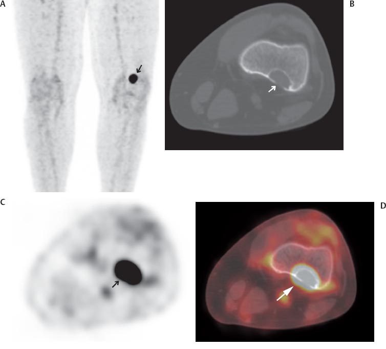
  2. In children, benign fibro-osseous lesions can mimic bone metastases on PET imaging. Furthermore, maximum SUVs of these benign lesions overlap those of malignant lesions. Information gained from correlative CT or MR images is useful in determining the benign nature of such lesions (Fig. 26.2).5,6
  3. Maximum SUVs of primary Ewing sarcoma and osteosarcoma may not accurately reflect overall tumor necrosis because small metabolically active foci may be present within tumor that is > 90% necrotic.3,7
  4. After chemotherapy or radiation therapy, SUVs may remain high within primary bone tumors due to inflammation or reactive fibrosis rather than viable tumor.3,7

image Soft Tissue Tumors


Clinical Indication

In children with soft tissue malignancies, PET/CT has shown value in identifying the site of an unknown primary tumor, staging, monitoring response to therapy, and detecting recurrence.


Accuracy


  1. Because soft tissue malignancies in children are rare, there have been no formal studies of the accuracy of PET or PET/CT in the diagnosis or follow-up of these tumors.

Sep 3, 2016 | Posted by in GENERAL RADIOLOGY | Comments Off on Pediatric PET/CT

Full access? Get Clinical Tree

Get Clinical Tree app for offline access