and Zdeněk Fryšák1
(1)
Department of Internal Medicine III – Nephrology, Rheumatology and Endocrinology, Faculty of Medicine and Dentistry, Palacky University Olomouc and University Hospital Olomouc, Olomouc, Czech Republic
Keywords
EthanolThyroid cystsParathyroid adenomaAlternative to surgery23.1 Method, Indications, and Complications of PEIT
Percutaneous ethanol injection therapy (PEIT) is a minimally invasive procedure that may be performed as an alternative to surgery for the treatment of thyroid cysts, parathyroid adenoma (PAd) and, less frequently, toxic thyroid nodules [1, 2]. Most recently, PEIT of metastatic lymph nodes is gaining interest as a nonsurgical directed therapy for patients with recurrent differentiated thyroid carcinoma (PTC and FTC) [3].
Absolute ethanol (96%) induces cellular dehydration, protein denaturation, and thrombosis in the capillary bed. This is followed by coagulation necrosis and reactive fibrosis in parathyroid adenoma or at the cyst wall, resulting in their shrinkage [4, 5].
PEIT is suitable procedure for elderly patients with comorbidities and high surgical risk. For anxious patients or those not willing to undergone surgery PEIT may be method of choice [2, 6].
We require FNAB before performing PEIT of large PAd (in small PAd <1 mL and in PAd with typical US features malignancy is not expected) or the thickened wall of a complex cyst, including the examination of evacuated fluid. PEIT is only performed if there is no suspicion of malignant lesion.
The risk of overlooking thyroid malignancy, including PTC and FTC microadenomas, in those treated with minimally invasive techniques exists, but must be very small. During long-term follow-up (> 5 years), no patient operated on due to growth and/or pressure symptoms has been diagnosed with thyroid malignancy after PEIT [7].
Complications can be caused by leakage of ethanol into the surrounding tissue [4, 5]:
No serious, life-threatening complications were reported in any published series of patients.
Mild localized pain for 24–48 h after application is typically noted.
Dysphonia or temporary vocal fold paresis; permanent paresis is rare; in the largest sample of 432 patients with thyroid cysts undergoing percutaneous ethanol injection therapy, temporary paresis was noted in 0.7% [8].
Small hematoma.
Periglandular fibrosis following repeated percutaneous ethanol injection therapy, complicating potential later surgery.
Frequency of PEIT complications is lower in thyroid cysts [5].
PEIT is relatively effective, often offering the same results as surgery. It is safe, inexpensive, may be repeated and carried out on outpatient basis.
PEIT is mostly performed without local anesthesia [5].
It must be stressed that PEIT is not a routine procedure and is only performed in selected patients. It should be carried out by a specialist routinely performing FNAB of thyroid nodules and PEIT.
23.2 Ultrasound-Guided Percutaneous Ethanol Injection Therapy (US-PEIT) of Thyroid Cysts
Simple aspiration of the cystic portion of the nodule can reduce pressure-related symptoms and cosmetic problems. Although aspiration can induce collapse of the cystic portion, there is a high risk of fluid recurrence 10% up to 80% [6, 10].
Previously, Treece et al. in 1983 or Edmonds et al. in 1987 successfully used instillation of intracystic tetracycline hydrochloride as sclerosant to treat recurrent pure thyroid cyst [12, 13].
PEIT of thyroid cyst was first successfully performed by Croatian physician Rozman in 1989 [14].
US-PEIT is indicated in patients with recurrent thyroid cysts with volumes ≥3 mL causing symptoms of compression or cosmetic complaints [6].
For the purpose of PEIT, cyst can be divided according to US pattern, size, and liquid character obtained by FNAB [5, 6, 9, 11]:
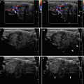
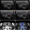
Pure cysts (Fig. 23.1aa)—US scan showing a cystic component of more than 90%, anechoic content, and smooth internal wall; drained fluid is viscous, clear, or pale yellow.
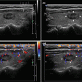
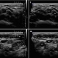
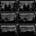
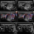
Complex cysts (Fig. 23.2bb, dd)—US scan showing a fluid component of 60–90% of the volume, anechoic or flocculated contents, roughened wall and septa in some cysts; aspirated fluid is mostly brown with debris, or dark yellow, viscous-to-gelatinous, or hemorrhagic.


Fig. 23.1
(aa) A 59-year-old woman with a 1 month palpable resistance on the neck. Solitary pure cyst (arrowheads), size 35 × 27 × 22 mm and volume 11 mL in the RL prior to PEIT: smooth wall; anechoic contents; Tvol 24 mL, RL 20 mL and LL 4 mL; transverse.(bb) Detail of pure cyst (arrowheads): smooth wall; anechoic contents; transverse. (cc) Detail of pure cyst (arrowheads): smooth wall; anechoic contents; longitudinal. (dd) Six months post PEIT—small solid nodule (arrows), size 10 × 8 × 6 mm and volume 0.3 mL as residue of cyst: inhomogeneous structure; mostly isoechoic, focally hypoechoic areas; no recurrence of the liquid component; Tvol 12 mL, RL 7 mL, and LL 5 mL; transverse. (ee) Detail of small solid residue (arrows): inhomogeneous structure; mostly isoechoic, focally hypoechoic areas; no recurrence of the liquid component; transverse. (ff) Detail of small solid residue (arrows): inhomogeneous structure; mostly isoechoic, focally hypoechoic areas; no recurrence of the liquid component; longitudinal


Fig. 23.2
(aa) A 63-year-old man with a 2 months progressively increasing, palpable resistance (full black line) on the right side of the neck. Personal history: myocardial infarction and cardiopulmonary resuscitation with tracheotomy–scar (long black arrow). US revealed giant complex cyst volume 102 mL in the RL. Patient is lying before evacuation with visible resistance, also visible scar post tracheotomy. (bb) Lying patient post evacuation and first PEIT, visible resistance (dotted black line) disappeared. Five filled 20 mL syringes with brownish content. (cc) Overall US view of giant complex cyst (arrowheads), occupying whole RL, size 76 × 63 × 41 mm, volume 102 mL and normal sized LL: smooth wall with short thick peripheral septa (open arrows); anechoic content; scar after tracheotomy (long arrows)—interrupting continuity of the isthmus and deformed trachea with indentation of the wall; Tvol 110 mL, asymmetry—RL 102 mL and LL 8 mL; transverse. (dd) Detail of giant complex cyst (arrowheads): smooth wall with short thick peripheral septa (open arrows); anechoic contents; transverse. (ee) Detail of giant complex cyst (arrowheads): smooth wall without septa; anechoic contents; longitudinal. (ff) Twelve months post PEIT—small solid nodule (arrows), size 15 × 11 × 9 mm and volume 0.3 mL as residue of cyst: solid; inhomogeneous; mostly hypoechoic; tiny bands and punctuations of fibrosis (open arrows); no recurrence of the liquid component; shrunken scar post tracheotomy (long arrows); Tvol 19 mL, RL 11 mL, and LL 8 mL; transverse. (gg) Detail of small solid residue (arrows): solid; inhomogeneous structure; mostly hypoechoic; thin bands and areas of fibrosis (open arrows); transverse. (hh) Detail of small solid residue (arrows): solid; inhomogeneous structure; mostly hypoechoic; thin bands and areas of fibrosis (open arrows); longitudinal
Regarding size, cysts are mostly divided into small (3–10 mL), medium (11–40 mL), and large (> 40 mL).
Criteria of complete therapeutic success [6]:
During one application, amount of instilled ethanol is equal to 23–100% of the initial cyst volume but preferably no more than 10 mL. Based on ultrasound findings, procedure is repeated at intervals of 2 weeks to 1 month [16, 17].
A special issue presents in viscous cysts with thick gelatinous content, which cannot be aspirated with an 18-gauge needle. These required different techniques:
Most commonly used is a two-stage ethanol ablation technique. During the first session, ethanol is injected into the cyst (1 mL for each 10 mL of cyst volume) to reduce density of the viscous fluid. At the second session (2–4 weeks after the first), the cystic fluid is aspirated from the nodule, and ethanol is injected [11, 18].
Sung et al. attempted a one-step ethanol ablation technique with thick gelatinous content aspiration through large-bore needle or a catheter connected to a suction pump following the ethanol injection [15].
Cystic fluid is aspirated as completely as possible and then ethanol is instilled into the cavity to a volume of 40–100% of the volume of aspirated fluid [5].
Evaluating more than 20 years of experiences, PEIT is considered in the United States and Europe by the 2010 AACE/AME/ETA Guidelines as a standard nonsurgical, minimally invasive procedure in management of recurrent thyroid cysts [19].
23.3 Ultrasound-Guided Percutaneous Ethanol Injection Therapy (US-PEIT) of the Parathyroid Gland
Another term often used for this method is Percutaneous Alcohol Ablation of the Parathyroid gland (PAAP) [20].
First, it should be noted that surgery is the gold standard and permanently effective therapy for patients with primary hyperparathyroidism (pHPT). Therapeutic effect of conventional exploration and minimally invasive parathyroidectomy for pHPT for both is about 97% [21].
Surgery is indicated in all symptomatic patients with PHPT.
Stay updated, free articles. Join our Telegram channel

Full access? Get Clinical Tree


