Location: Mainly in the limbs: distal femur, proximal tibia, and proximal humerus.
Clinical: Swelling with little or no pain.
Imaging: On x-ray – globose mass lying on the outer surface of the cortex, very radiolucent or with granules, rings, spots of cartilage, and rarely bunches of faded ossifications. Outer cortical erosion, saucerlike cortex, thickness of the adjacent cortex, buttress of periosteal reaction, internal sclerotic rim, and sharp margins. On CT – confirms periosteal site of the tumor without involvement of the medullary canal.
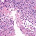
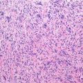
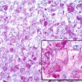
Histopathology: Like peripheral C, it is well differentiated. Rarely myxoid. Grade 1 is considered a periosteal chondroma, grade 2 is frequent, and grade 3 is rare.
Course and Staging: Rare recurrence if surgery is adequate; late, very rare metastasis. Usually stage IA.
Treatment: Wide resection. Prognosis is good.
Key Points
Clinical | Adults, swelling |
Radiological | Subperiosteal, metaphyseal, with erosion of the cortex, granular calcifications, periosteal reaction |
Histological | Lobules of low-grade cartilage |
Differential diagnosis
Stay updated, free articles. Join our Telegram channel
Full access? Get Clinical Tree
 Get Clinical Tree app for offline access

|


