Lower Extremity Arterial Duplex Ultrasonography Overview
Location
• Abdominal aorta: originates at the aortic hiatus of the diaphragm, courses left of midline, anterior to the vertebrae, and terminates at the fourth lumbar vertebra.
• Aortic bifurcation: originates approximately at level of the umbilicus to form the common iliac arteries.
• Common iliac arteries: terminate at the origin of the internal iliac (hypogastric) and external iliac arteries.
• External iliac arteries: originate at the common iliac artery bifurcation; form the common femoral artery just proximal to the inguinal ligament.
• Lower extremity arterial system: originates with common femoral artery just inferior to the inguinal ligament.
Anatomy
• Abdominal aorta: lies to the left of the midline and courses along the lower abdomen with a slight posterior-to-anterior angulation. The aorta tapers as it moves toward its bifurcation where it has a diameter of approximately 1.5 cm in a normal adult. It gives rise to the iliac arteries, which course through the pelvis to form the common femoral artery at the level of the inguinal ligament.
• Common femoral artery: lies lateral to the common femoral vein and bifurcates into the profunda femoris (deep femoral) artery and the superficial femoral artery.
• Profunda femoris artery: lies lateral to the superficial femoral artery and gives rise to branches that supply the region of the femoral head and the deep thigh muscles. In a small percentage of patients, the profunda femoris will originate medially from the common femoral artery.
• Superficial femoral artery: lies anterior to the femoral vein and courses along the anteromedial aspect of the thigh. It courses deep as it enters the adductor canal in the distal thigh where it becomes the popliteal artery.
• Popliteal artery: within the popliteal fossa lies anterior and medial to the popliteal vein and gives rise to small geniculate branches. It branches into the anterior tibial artery and the tibioperoneal trunk.
• Anterior tibial artery: passes superficial to the interosseous membrane and courses anteriorly along the membrane. Distally, the anterior tibial artery courses along the anterior aspect of the tibia and becomes the dorsalis pedis artery after crossing the ankle joint.
• Tibioperoneal trunk: gives rise to the posterior tibial and peroneal arteries that supply the calf muscles.
• Posterior tibial artery: originates between the tibia and the fibula and courses along the medial aspect of the calf to a midpoint between the medial malleolus (ankle bone) and the heel.
• Peroneal artery: originates from the tibioperoneal trunk and courses obliquely along the medial aspect of the fibula. The peroneal artery lies deep to the posterior tibial artery and in close apposition to the fibula.
Physiology
• The lower extremity arteries supply blood to the muscles of the thigh and calf. Normal resting muscle has a low flow demand. In contrast exercising muscle requires a significant increase in flow volume to oxygenate the tissues and carry away exercise-related toxins.
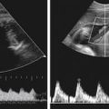
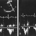
• In the resting state, normal muscle will exhibit a high-resistance blood flow pattern characterized by low diastolic flow. In contrast, exercising muscle has a low-resistance blood flow pattern, exhibiting constant forward diastolic flow.
Sonographic Appearance
• The normal vessel lumen should be echo-free.
• Using multiple scan planes, you should be able to image the vessel bifurcations, which may be off-axis.
Preparation
Patient Prep
• None.
Pulsed Doppler Transducers
• 5.0-MHz to 7.5-MHz linear array for thigh and calf in majority of patients.
• 2.0-MHz to 4.0-MHz curved array for abdominal aorta and iliac arteries and may be required to image upper thigh vessels in patients of large body habitus.
Patient Position
• Beginning with the patient in a supine position, obtain bilateral brachial systolic blood pressures using an appropriately sized blood pressure cuff (the width of the cuff bladder should exceed 20% of the limb diameter) and a continuous-wave Doppler transducer. Record the pressures on the worksheet (see the section on Data Documentation: Using Blood Pressures). In a similar manner, obtain bilateral ankle systolic pressure measurements from the dorsalis pedis and posterior tibial arteries by placing an appropriately sized blood pressure cuff immediately above the ankle. Record all tibial pressures on the worksheet and calculate the ankle/brachial index for both legs.
• Continue with the patient lying supine with the leg rotated outward and the knee slightly flexed in a frog-leg position to allow access to the common femoral artery and the popliteal fossa.
• Or have the patient lie prone with the feet slightly elevated on a pillow or rolled towel to allow access to the popliteal fossa.
• You may stand beside the examination table to scan the limb nearest to you. To ensure appropriate ergonomic positioning, make sure that your arm is supported and not at a right angle to your body and that the examination table has been raised to a height that is comfortable for you.
Lower Extremity Arterial Duplex Survey
1. The purpose of the survey is to identify arterial wall abnormalities, vessel tortuosity, pathology, and anatomic anomalies. The initial survey is performed with gray-scale (B-Mode) imaging to ensure that features of the arterial wall and plaque morphology are accurately characterized.
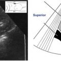
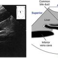
2. As clinically indicated or requested, initiate the survey superior to the umbilicus to image the mid-to-distal abdominal aorta. Beginning with the transducer in a transverse imaging plane, locate the axial section of the inferior vena cava lying to the right of the abdominal midline. Move the transducer left lateral to image the aorta.
3. Begin the axial survey of the mid-to-distal abdominal aorta. Scan inferiorly, noting vessel walls, tortuosity, diameter changes, and anatomic anomalies.

4. Locate the aortic bifurcation and identify the right and left common iliac arteries. Make special note of the presence of plaque and/or aneurysmal dilation.
5. Continue to scan inferiorly along the course of the right common iliac artery, noting its bifurcation into the internal and external iliac arteries and the curving deep to superficial course of the external iliac artery. The internal iliac artery commonly lies deep to the external iliac artery. The external iliac artery lies superior to the external iliac vein.
Images in this chapter courtesy of Penn State Hershey Vascular Noninvasive Diagnostic Laboratory, Hershey, Pa.
6. Continue to scan inferiorly to follow the external iliac artery to the level of the inguinal ligament where it becomes the common femoral artery.
7. Immediately inferior to the inguinal ligament, confirm the identity of the common femoral artery by identifying the common femoral vein. If the iliac arteries have been scanned in the sagittal plane, rotate the transducer 90 degrees to assume a transverse imaging plane. Apply firm compression of the tissues over the vein with the transducer held perpendicular to the anterior vessel wall to distinguish the compressible vein from the noncompressible artery (see the section on Peripheral Venous Duplex Sonography in this chapter).

8. Move the transducer laterally to identify the common femoral artery.
9. Continue the axial survey of the right common femoral artery. Angle as superiorly as possible to image the proximal right common femoral artery.
10. Scan inferiorly, noting vessel walls, tortuosity, and anatomic anomalies.
11. At the level of the bifurcation, make special note of the presence of plaque and the origin of the bifurcation vessels.
12. Scan the proximal to mid profunda femoris artery.
13. Return to the common femoral artery bifurcation.
14. Continue to follow the course of the superficial femoral artery, imaging as far inferiorly as possible.
15. At the level of the distal thigh, in the region of the adductor canal, the superficial femoral artery courses deeper into the tissues to become the popliteal artery. Visibility of the artery may be compromised at this level of the limb.
16. Move the transducer behind the knee into the popliteal fossa. With your free hand placed over the patella (knee cap), apply counterpressure to the knee. Angle the transducer superiorly and slightly medially to image the popliteal artery and vein. Confirm the identity of the artery by using a venous compression maneuver. Scan as superiorly into the distal thigh as possible.
17. Continue to follow the course of the popliteal artery, noting the presence of small geniculate branches and any diameter changes that would signify aneurysmal dilation.
18. Approximately 6 to 8 cm below the knee, the popliteal will give rise to the anterior tibial artery and the tibioperoneal trunk.
19. Follow the course of the proximal anterior tibial artery, noting the presence of plaque.
20. Return to the popliteal artery and follow the course of the tibioperoneal trunk. Note its bifurcation into the posterior tibial artery and the peroneal artery.
21. Follow the more superficial posterior tibial artery to the level of the medial malleolus (ankle) as it courses along the medial aspect of the calf.
22. Return to the bifurcation of the tibioperoneal trunk.
23. Follow the deeper lying peroneal artery to the level of the ankle as it courses along the medial aspect of the calf.
24. Return to the level of the umbilicus to complete the survey of the mid-to-distal abdominal aorta in the sagittal imaging plane.
25. Locate the inferior vena cava and the abdominal aorta. Rotate the transducer 90 degrees into the sagittal scanning plane to obtain a longitudinal view of these vessels.
26. Follow the course of the mid-to-distal aorta to the level of the bifurcation into the common iliac arteries. Obtain appropriately angle-corrected (angle of insonation 60 degrees or less with the cursor parallel to the vessel wall) Doppler spectral waveforms from the distal aorta, making careful note of regions of flow disturbance or turbulence.
27. Identify the right common iliac artery. Follow the longitudinal course of this vessel in the sagittal imaging plane. Obtain representative angle-corrected Doppler spectral waveforms from this vessel segment.

28. Continue to follow the course of the common iliac artery into the external iliac artery. Obtain representative Doppler spectral waveforms from this vessel segment.
29. Continue to move inferiorly to identify the longitudinal section of the common femoral artery. Obtain representative Doppler spectral waveforms from this vessel segment.
30. Identify the common femoral artery bifurcation. Scan the proximal to midsegment of the profunda femoris artery. Obtain representative Doppler spectral waveforms from this vessel segment.

31. Return to the common femoral artery bifurcation. Identify the origin of the superficial femoral artery. Obtain longitudinal images of this vessel segment and record representative Doppler spectral waveforms.
32. Continue to follow the course of the superficial femoral artery. Obtain longitudinal images of the mid and distal segments and record representative Doppler spectral waveforms from these vessel segments.
33. Place the transducer in the popliteal fossa to obtain longitudinal images of the popliteal artery. Obtain representative Doppler spectral waveforms from this vessel segment.

34. Locate the proximal anterior tibial artery. Obtain longitudinal images of the vessel origin and proximal segment. Record representative Doppler spectral waveforms from this vessel segment.

35. Return to the popliteal artery and identify the tibioperoneal trunk. Obtain longitudinal images of the origin and proximal segment of the peroneal artery. Record representative Doppler spectral waveforms from this vessel segment.

36. Continue to follow the course of the peroneal artery in the sagittal imaging plane. Obtain images of the mid and distal segments of the artery and record representative Doppler spectral waveforms from these vessel segments.
37. Return to the popliteal artery and identify the tibioperoneal trunk. Obtain longitudinal images of the origin and proximal segment of the posterior tibial artery. Record representative Doppler spectral waveforms from this vessel segment.
38. Continue to follow the course of the longitudinal section of the posterior tibial artery in the sagittal imaging plane. Obtain images of the mid and distal segments of the artery and record representative Doppler spectral waveforms from these vessel segments.
39. Repeat the survey on the contralateral limb.
Lower Limb Arterial Duplex Required Images


12. Doppler spectral waveform from the PROXIMAL SUPERFICIAL FEMORAL ARTERY.

13. Longitudinal image of the MID SUPERFICIAL FEMORAL ARTERY.
Related posts:
Stay updated, free articles. Join our Telegram channel

Full access? Get Clinical Tree


