18 Peripheral Nerves
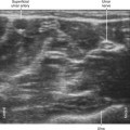
Peripheral nerves have a fascicular or “honeycomb” echotexture. This consists of the mixture of nerve fiber (hypoechoic) and connective tissue (hyperechoic) content within the nerve. Because there is little connective tissue within more central nerves (e.g., the cervical ventral rami of the brachial plexus), these nerves have a monofascicular or oligofascicular appearance on ultrasound scans.1 Nerves that are surrounded by hypoechoic muscle are usually easier to visualize than nerves that are surrounded by hyperechoic fat because the nerve borders are more evident.
Peripheral nerves have a complex architecture. Nerves are like a plexus within themselves, with fascicles combining and recombining internally along the nerve path. Because of this intertwined network, the fascicle count varies along the nerve path. Nerve sections taken 2 mm apart can have different fascicular patterns.2 The connective tissue content and fascicle count of peripheral nerves vary directly. That is, the amount of connective tissue is more abundant in multifascicular nerves.3 The connective tissue within nerves protects the fascicles from injury. Therefore, monofascicular nerves are more vulnerable to damage.
Identification of nerve fascicles is the basis of peripheral nerve imaging. With ultrasound, only a small subset of the total number of fascicles is imaged. In one study of ex vivo nerve specimens, only about one third the number of fascicles visible on light microscopy were visible on ultrasound scans.4 It is difficult for imaging technologies to resolve thin collagenous boundaries between adjacent fascicles. For these reasons, fascicular discrimination is a standard by which to judge nerve imaging quality.
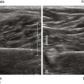
High ultrasound frequencies (10-15 MHz) provide better resolution of nerve fascicles.5 However, at lower frequencies (7-10 MHz), peripheral nerves are still visible as cordlike structures.5 Commercial nerve imaging presets of imaging quality controls have been developed that enhance detection of nerve fascicles.
Related posts:


Stay updated, free articles. Join our Telegram channel

Full access? Get Clinical Tree


