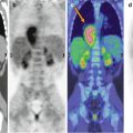
Fig. 15.1
A 14-year-old girl treated for Hodgkin’s lymphoma. Maximum intensity projection (a) and axial CT and PET/CT fusion images (b) show moderately intense FDG uptake in the right breast, corresponding to phylloid tumor (yellow arrow in b)
References
1.
Abdallah A, Saklaoui O, Stückle C, Sommerer F, Hatzmann W, Audretsch W, Wesemann A, Zink M, Skoljarev L, Papadopoulos S (2009) Case reports of operative management of very large, benign phylloid tumors – is a safety margin necessary? Gynakol Geburtshilfliche Rundsch 49:320–325PubMedCrossRef
Related posts:


Stay updated, free articles. Join our Telegram channel

Full access? Get Clinical Tree


