Fig. 1.1
Chest radiograph shows the presence of a radiopaque FB (coin) lodged between the hypopharynx and the cervical esophagus
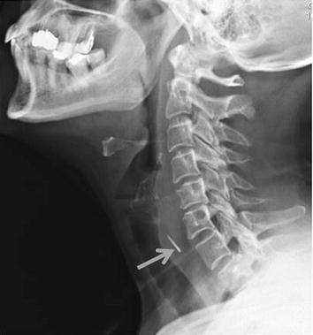
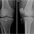
Fig. 1.2
Neck lateral plain film shows the presence of a radiopaque FB (fish bone, arrow) anterior to the body of C6
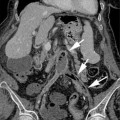
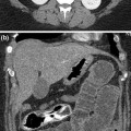
Multidetector row computed tomography (MDCT) is very useful for the evaluation of patients with suspected pharyngoesophageal FBs (Fig. 1.3) because it offers short examination time and the ability to obtain diagnostically useful coronal and sagittal reconstruction images. MDCT can detect ingested objects such as slightly calcified objects that are missed by conventional radiographs. Moreover, MDCT is also helpful to detect FB complications (Fig. 1.4), such as perforation, fistula, or abscess [9].
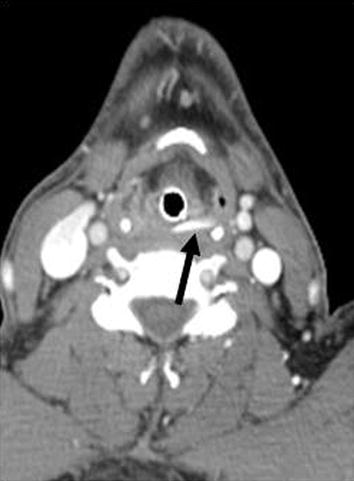
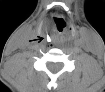
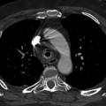
Fig. 1.3
MDCT shows the presence of an impacted hypopharyngeal FB (fish bone, arrow)
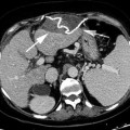
Fig. 1.4
MDCT demonstrates an impacted hyperdense FB (arrow) at the level of the hypopharynx with a contiguous abscess
1.5 Neck Soft Tissue Foreign Bodies
A retained FB in the soft tissue of the neck (Fig. 1.5) may determine severe infection or inflammatory reaction: due to this reason, the detection and the removal of the FB is necessary [13].
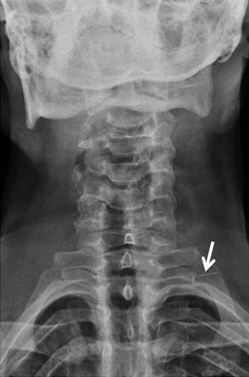
Fig. 1.5
Neck anteroposterior radiograph shows the presence of a linear radio-opaque FB (needle, arrow) located in the soft tissues
Bullet injuries can determine the presence of a FB in the soft tissue (Fig. 1.6). Bullets are usually described by their caliber, which is a measurement of their diameter in inches or in millimeters. Although the caliber of a bullet is important, it has only a loose relationship to the weight of the bullet and the size of its charge. These latter parameters help determine the kinetic energy of the bullet, which is an important factor in determining its wounding potential. Bullet injuries are most severe in friable solid organs, where damage may be caused by temporary cavitation remote from the actual bullet track. Dense tissues (e.g., bone) and loose tissues (e.g., subcutaneous fat) are more resistant to bullet injury. Bones modify the behavior of bullets markedly, altering their course, slowing them down, and increasing their deformity and fragmentation [14].
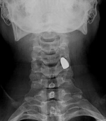
Fig. 1.6
Radiograph of the neck shows the presence of a projectile lodged in the soft tissues
Imaging techniques are required to identify the FB and establish its exact location prior to surgical removal attempt. Radiographic evaluations are routinely performed in order to confirm the presence of radio-opaque foreign bodies in the soft tissues of the neck such as glass, metal, and stone within the soft tissue [15, 16].
Limitations of radiography include non-visualization of radiolucent foreign bodies, radiation exposure and failure of precise localization during removal.
Radioscopy offers a more accurate topographic assessment and allows reference points to be marked on the skin to aid subsequent FB removal. However, radioscopy exposes patient and operator alike to relatively high doses of ionizing radiation.
Related posts:
Stay updated, free articles. Join our Telegram channel

Full access? Get Clinical Tree


