• Postulated to arise from ossification of lateral segment of posterior atlantooccipital ligament or joint capsule
• Morphologically normal bone
Clinical Issues
• Most common: Asymptomatic
• Other symptoms
Vertebrobasilar ischemia or infarction
Vertigo
Headache
Neck pain
Diagnostic Checklist
• Vertebral arteries predisposed to surgical injury during lateral mass screw placement
• Multiplanar bone CT with 3D reformats best demonstrates ponticulus posticus
(Left) Lateral radiograph of the upper cervical spine demonstrates a partial osseous roof over the C1 vertebral artery foramen, characteristic of ponticulus posticus (incomplete variant).
(Right) Lateral radiography of the upper cervical spine reveals a complete osseous roof over the C1 vertebral artery foramen, typical of classic ponticulus posticus (complete variant).
(Left) Sagittal CTA depicts a complete C1 arch, with a robust osseous covering (complete ponticulus posticus) over the vertebral artery along the superior aspect of the C1 arch.
(Right) Anteroposterior view of a CTA 3D reformat shows a dominant left vertebral artery and smaller (nondominant) right vertebral artery . Both are covered by a complete osseous bridge along the superior aspect of the C1 arch.


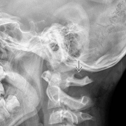
 over the C1 vertebral artery foramen, characteristic of ponticulus posticus (incomplete variant).
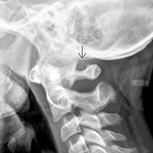
 over the C1 vertebral artery foramen, typical of classic ponticulus posticus (complete variant).
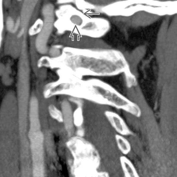
 over the vertebral artery
  along the superior aspect of the C1 arch.
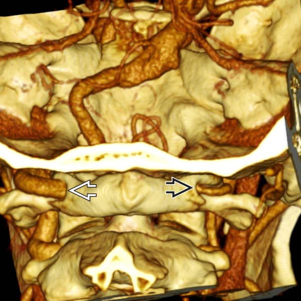
 and smaller (nondominant) right vertebral artery
  . Both are covered by a complete osseous bridge along the superior aspect of the C1 arch.