Site of presentation may depend partially on transplanted organ
Extranodal (80%) > nodal involvement (20%)
• Imaging findings of post-transplant lymphoproliferative disorder parallel those of non-Hodgkin lymphoma (NHL) in immunocompetent patients
• GI tract: Imaging findings are similar to NHL, including mass-like bowel wall thickening, aneurysmal dilatation, ulcerated polyploid mass, or submucosal nodules
Increased prevalence of ulceration and bowel perforation
• Liver: Most frequently involved abdominal solid organ
Single or multiple poorly enhancing masses, discrete mass in porta hepatis, or diffuse infiltration of liver
• Spleen: Splenomegaly ± discrete lesions (usually multiple, hypoattenuating, and variable in size)
• Kidney: Most common site in renal transplant recipients
Heterogeneous mass surrounding hilar vessels, parenchymal masses, or diffuse infiltrative disease
• Nodal disease: Abdominal nodal involvement in only 15-20% of cases
Nodal involvement much less common than in immunocompetent NHL
TOP DIFFERENTIAL DIAGNOSES
• Recurrent or new malignancy
• Opportunistic infections
PATHOLOGY
• Most cases are related to B-lymphocyte proliferation due to Epstein-Barr virus (EBV) infection
CLINICAL ISSUES
• High mortality, with survival rates of only 25-35%
• Treatment: Reduction or cessation of immunosuppression can be effective, although antiviral drugs, chemotherapy, or rituximab may be necessary
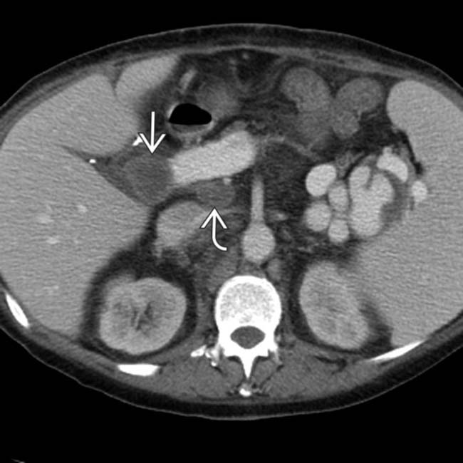
(Left) Axial CECT in a patient post liver transplant demonstrates a new hypodense mass in the porta hepatis, as well as an enlarging portacaval lymph node .
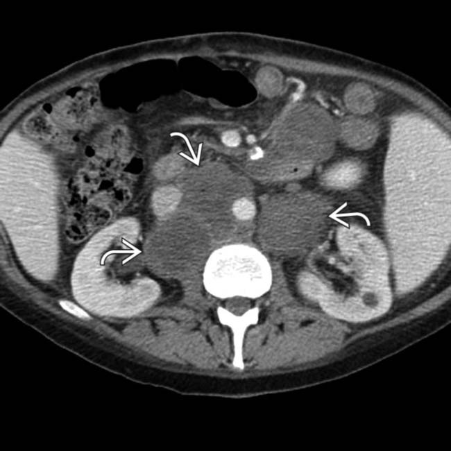
(Right) Axial CECT in the same patient demonstrates extensive retroperitoneal lymphadenopathy . The findings of post-transplant lymphoproliferative disorder (PTLD) in this case are indistinguishable from traditional non-Hodgkin lymphoma (NHL) in an immunocompetent patient.
(Left) Axial NECT demonstrates mass-like wall thickening of a segment of colon with aneurysmal dilatation.
(Right) Coronal NECT in the same patient again demonstrates the significant wall thickening of the bowel segment with dilatation. This is a common appearance for both NHL in immunocompetent patients and PTLD.
• Heterogeneous group of lymphoproliferative diseases that occur in post-transplant setting (either solid organ or stem cell transplants), ranging from abnormal lymphoid hyperplasias to frank malignancies
IMAGING
General Features
• Location
Extranodal involvement (80%) is much more common than nodal involvement (20%)
– Unlike lymphoma in general population where nodal disease predominates
Can occur nearly anywhere, with common locations including lungs, GI tract, and CNS
– Site of presentation may depend partially on type of transplanted organ
– Abdominal cavity is most frequently involved (up to 50% of all cases)
– May occur within renal and liver allografts
Some studies have suggested that PTLD may preferentially affect allograft itself
• Size
Masses and nodes range from < 1 cm to huge masses
Imaging Recommendations
• Best imaging tool
CECT for initial diagnosis
PET/CT for staging and follow-up
CT Findings
• Imaging findings of PTLD mostly parallel those of non-Hodgkin lymphoma (NHL) in immunocompetent patients


 in the porta hepatis, as well as an enlarging portacaval lymph node
  .
.
 . The findings of post-transplant lymphoproliferative disorder (PTLD) in this case are indistinguishable from traditional non-Hodgkin lymphoma (NHL) in an immunocompetent patient.
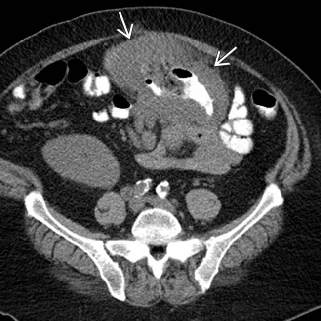
 of a segment of colon with aneurysmal dilatation.
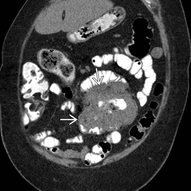
 of the bowel segment with dilatation. This is a common appearance for both NHL in immunocompetent patients and PTLD.


 Most commonly involved site in renal transplant recipients and may affect native kidneys or allograft


