4 Posterior Intercostal Arteries
D. Hortung, K. Hueper
The posterior intercostal arteries III to XI, the subcostal artery, and the lumbar arteries remain segmental posterior branches of the descending aorta.1,2 When the heart descends, it causes the more cranial intercostal arteries to take an upward course. Their origins are therefore near to each other, explaining the occurrence of trunk formations of two or more arteries, especially of the third and fourth arteries. During embryologic development, a longitudinal anastomosis is formed between the segmental arteries C7, T1, and T2 and the original connections to the aorta disappear, resulting in the costocervical trunk. As a rule the anastomosis runs anterior to the ribs, but sometimes it is positioned between the rib and the transverse process, like the vertebral artery, which runs through the transverse foramina in the cervical vertebral column.1–6
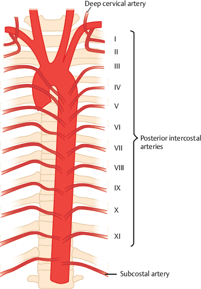
Fig. 4.1 “Normal” situation as given in textbooks. Schematic I–XI, posterior intercostal arteries.
4.1 Posterior Intercostal Arteries III–IX and Subcostal Artery

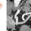
Fig. 4.2 Separate origins of the intercostal arteries of one segment (83%). Schematic (a) and MRA (b,c). MIP, transverse view (b) and VR 3D image, posterior view (c). 1 Right renal artery; 2 celiac trunk; 3 left intercostal artery; 4 right intercostal artery.

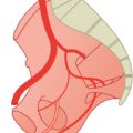
Fig. 4.3 Trunk formation of the intercostal arteries of one segment (2%). Schematic.
Related posts:


Stay updated, free articles. Join our Telegram channel

Full access? Get Clinical Tree


