Fig. 22.1
90° Propeller flap
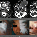
The patient suffered an open tibial and fibula pilon fracture (Gustilo IIIB) in a traffic accident. A propeller perforator flap based on the perforator vessels in the front of the leg was planned
In a preoperative perforator examination, several perforator vessels from the peroneal and anterior tibial artery were confirmed using MDCTA and Doppler sonography. A skin flap harvest was planned on the muscular fascia to match the area of skin loss (a) After ICG contrast angiography was performed to ensure flap flexibility, an aggressive perforator dissection was performed, thereby resulting in two dominant perforator vessels (b). The flap was then turned 90°. The flap survived completely (c)
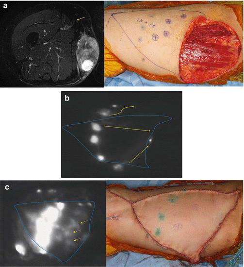
Fig. 22.2
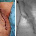
V-Y advancement flap
A wide defect persisted on the right thigh after sarcoma resection. Several perforator vessels were confirmed around the area of skin loss by MRA and Doppler sonography (a). The V-Y advancement perforator flap was designed in the direction that would not inhibit the lymph style and where skin tension did not appear. ICG contrast was injected subcutaneously to confirm the lymph style (b) ICG fluorescence angiography was performed to identify the blood vessels after perivascular detachment under the muscular fasciae, and several dominant perforator vessels were chosen (c). The flexibility of the skin flap was ensured, the flap survived completely, and lymphedema did not develop in the leg

Fig. 22.3
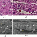
180° Propeller flap
The plate was exposed after bone fixation was performed around the left elbow. Two perforator vessels in the upper arm were identified preoperatively by MDCTA and Doppler sonography (a). The skin flap was cut circumferentially under the muscular fasciae to the area of the perforator vessels. After ICG fluorescence angiography was performed to confirm the run of the perivascular branch to penetrate the island skin flap using the Photodynamic Eye (b), the perforators were skeletonized to free it from adhesions and turned 180° to cover the loss region. The flap survived completely (c)
22.2.4.2 Free Perforator Flap
Free perforator flaps featuring vessel dissection, such as the ALT and DIEP flaps, have been reported to not be subjected to ICG fluorescent contrast examination. We often use this method at the time of the perforasome flap and perforator vessel confirmation during flap defatting (Fig. 22.4).
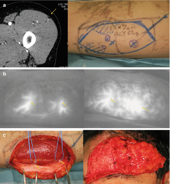

Fig. 22.4
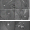
Free ALT flap
Frontal depression was performed after craniotomy. We planned the free ALT flap to include two perforator vessels. The perforator vessels were preoperatively confirmed using MDCTA and Doppler sonography (a). Intraoperative ICG fluorescence angiography was performed to confirm the perforator vessels and perforasome in the ALT flap (b). Defatting and thinning of the ALT flap were then performed (c). The flap survived completely
References
1.
2.
Saint-Cyr M, Schaverien M, Rohrich R (2009) Perforator flaps: history, controversies, physiology, anatomy, and use in reconstruction. Plast Reconstr Surg 123:132e–145eCrossRefPubMed
Stay updated, free articles. Join our Telegram channel

Full access? Get Clinical Tree


