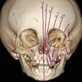
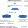
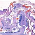
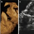
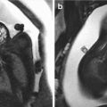
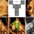
Fig. 20.1
Two- and three-dimensional ultrasound using surface-rendering and “skeleton” mode showing increased nuchal fold, prenasal edema, femoral hypoplasia, curved tibia and absent fibula, and oligodactyly of hands and feet associated with syndactyly of the right hand (arrows). The fetal face was characterized by a Tessier type 7 cleft (macrostomia) extending from the left commissure of the mouth and pointing to the chin with sunken appearance of the cheek (see autopsy finding for comparison) (arrow)
A presumptive prenatal diagnosis of Tessier number 7 cleft (macrostomia) associated with femoral hypoplasia-unusual facies syndrome (FH-UFS) was considered. Due to multiple severe congenital malformations, termination of pregnancy was performed using vaginal administration of PGE. Postmortem examination confirmed the antenatal diagnosis.
20.3 Discussion
FH-UFS (OMIM 134780) is a rare and sporadic skeletal disorder that was first described by Daentl in 1975 [13]. Nowaczyk et al. [14] emphasized the role of 3D ultrasound in the early prenatal diagnosis of femoral facies syndrome detected at 12, 15, and 19 weeks of gestation, respectively.
Tessier [2] classified facial clefts below the orbit and numbered them from 0 to 7, where number 7 represents a lateral cleft. Lateral facial clefting or macrostomia is an atypical cleft that represents about 3.1 % of all clefts. It may arise due to failed penetration of ectomesenchyme between the developing maxillary and mandibular prominences. It can be seen as an isolated defect or associated with features consistent with skeletal dysplasia [12]. The incidence is estimated as 1 in 50,000–175,000 live births [2]. The unilateral type is six times more frequently observed than the bilateral type [2]. Lampert [15] and Robinow et al. [6] demonstrated an autosomal dominance inheritance. Presti et al. [12] and Pilu et al. [11] reported the prenatal ultrasound detection of bilateral and unilateral Tessier number 7 cleft at 26 and 22 weeks of gestation, respectively. Lateral facial clefting may be part of an overlapping genetic syndrome such as mandibulofacial dysostosis and oculo-auriculo-vertebral spectrum [17, 18].
References
1.
2.
3.
Suzuki K, Hu D, Bustos T, Zlotogora J, Richieri-Costa A, Helms JA, Spritz RA. Mutations of PVRL1, encoding a cell-cell adhesion molecule/herpesvirus receptor, in cleft lip/palate-ectodermal dysplasia. Nat Genet. 2000;25:427–30.CrossRefPubMed
Stay updated, free articles. Join our Telegram channel

Full access? Get Clinical Tree


