A nucleus that undergoes ‘beta minus decay’ emits an electron (β-). This forms the basis of many nuclear medicine therapies. A nucleus that undergoes ‘beta plus decay’ emits a positron (β+) which undergoes annihilation when striking an electron (e-) to emit gamma rays travelling in opposite directions. This forms the basis of PET imaging. Changes in atomic number (Z) and mass number (A) are depicted
49.2 Gamma decay
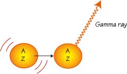
An excited nucleus that undergoes ‘gamma decay’ emits a gamma ray and therefore transitions to a more stable state. This forms the basis of most nuclear imaging techniques.
49.3 Gamma camera
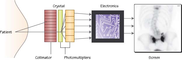
Gamma rays emitted from the patient are collimated to ensure the rays arrive at the crystal in a straight line. The crystal then converts these into photons. The photons are multiplied and converted to electrons which are then converted into an image
Fundamentals of nuclear medicine
Related posts:


Stay updated, free articles. Join our Telegram channel

Full access? Get Clinical Tree


