Pulmonary and Thoracic Lymphatic System
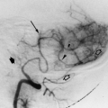
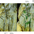
The pleural lymphatics are variable in size, number, and distribution. They distribute in a plexus with broad channels. There are abundant anastomoses with the pulmonary lymphatics. The lymphatic network is more prominent over the lower than the upper pulmonary lobes. The pleural lymphatics drain to the medial aspect of the lung near the hilum, where there are anastomoses with the lymphatics of the parenchymal plexus (Figs. 12.1, 12.2).
In the lungs, the lymphatic channels form two major paths: the first in the bronchoarterial and the other in the interlobular septal connective tissue. In both systems, the lymph flows toward the hilum, reaching the bronchial and mediastinal lymph nodes. Multiple anastomoses connect the interlobular perivenous lymphatics with those in the bronchoarterial sheathing. Anastomoses also exist among the bronchoarterial and pleural plexuses. The bronchoarterial lymphatics begin in the region of the distal respiratory bronchioles (Fig. 12.1).
Lymphatics of the Lungs and Pleura
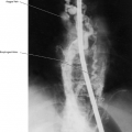
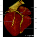
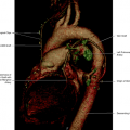
The lungs are subdivided into three main lymphatic drainage areas: superior, middle, and inferior, without correspondence to the pulmonary lobes. On the right lung, the superior area drains directly into the paratracheal and upper bronchopulmonary nodes. The middle zone drains into the paratracheal bifurcation and the central group of bronchopulmonary lymph nodes. The inferior zone drains into the inferior bronchopulmonary and bifurcation nodes and the posterior mediastinal chain. The right lymphatic duct is, therefore, the main drainage system of the right lung. On the left lung, the left superior area drains into the prevascular group of anterior mediastinal nodes and the left paratracheal nodes. The middle zone drains through the bifurcation and the central group of bronchopulmonary nodes and also directly through the paratracheal group. The inferior zone drains into the bifurcation and inferior bronchopulmonary nodes and into the posterior mediastinal group. The left lung, therefore, drains lymph from the superior zone and part of the middle zone to the left paratracheal nodes and into the thoracic duct. The remainder of lymph drainage of the left lung ends up in the right lymphatic duct (Fig. 12.3).
Thoracic Duct and Right Lymphatic Duct
The right lymphatic duct drains the great majority of lymph from both lungs, whereas the thoracic duct drains the apical portion of the left lung (Fig. 12.3).
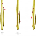
The thoracic duct originates at the cisterna chyli on the anterior aspect of the vertebral column at the lever T12 to L2, resulting from the junction of the lumbar lymphatic trunks. The thoracic duct enters the posterior mediastinum through the aortic hiatus of the diaphragm. The thoracic duct ascends cephalad at the right side of the aorta, approximately at the midline or slightly toward the right side. High up in the thorax the thoracic duct crosses to the left and leaves the thorax between the esophagus and left subclavian artery, joining the left subclavian vein from the posterior aspect. The diameter of the thoracic duct ranges from 1 to 7 mm, and valves are found in the majority of cases.
Related posts:
Stay updated, free articles. Join our Telegram channel

Full access? Get Clinical Tree


